


The crystal structure of RosB gives insights into the reaction mechanism of this first known member of a new family of flavodoxin-like enzymes
Konjik, V., Bruenle, S., Demmer, U., Vanselow, A., Sandhoff, R., Mack, M., Ermler, U.To be published.
Experimental Data Snapshot
Entity ID: 1 | |||||
|---|---|---|---|---|---|
| Molecule | Chains | Sequence Length | Organism | Details | Image |
| BRAMP domain protein | 257 | Streptomyces davaonensis | Mutation(s): 0 Gene Names: BN159_7989 | 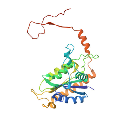 | |
UniProt | |||||
Find proteins for K4REZ6 (Streptomyces davaonensis (strain DSM 101723 / JCM 4913 / KCC S-0913 / 768)) Explore K4REZ6 Go to UniProtKB: K4REZ6 | |||||
Entity Groups | |||||
| Sequence Clusters | 30% Identity50% Identity70% Identity90% Identity95% Identity100% Identity | ||||
| UniProt Group | K4REZ6 | ||||
Sequence AnnotationsExpand | |||||
| |||||
| Ligands 1 Unique | |||||
|---|---|---|---|---|---|
| ID | Chains | Name / Formula / InChI Key | 2D Diagram | 3D Interactions | |
| 8DP Query on 8DP | I [auth A] J [auth B] K [auth C] L [auth D] M [auth E] | 8-demethyl-8-aminoriboflavin-5'-phosphate C16 H19 N5 O9 P SDTLZDHOYFRAPQ-WCQYABFASA-O |  | ||
| Length ( Å ) | Angle ( ˚ ) |
|---|---|
| a = 131.49 | α = 90 |
| b = 71.13 | β = 92.08 |
| c = 214.8 | γ = 90 |
| Software Name | Purpose |
|---|---|
| PHENIX | refinement |
| XDS | data reduction |
| XDS | data scaling |
| PHENIX | phasing |
RCSB PDB (citation) is hosted by
RCSB PDB is a member of the

