5MJ7

Structure of the C. elegans nucleoside hydrolase


Experimental Data Snapshot

  • Method: X-RAY DIFFRACTION
  • Resolution: 1.65 Å
  • R-Value Free: 0.154 
  • R-Value Work: 0.137 
  • R-Value Observed: 0.138 

wwPDB Validation   3D Report Full Report


This is version 1.3 of the entry. See complete history


Literature

Structural and biochemical characterization of the nucleoside hydrolase from C. elegans reveals the role of two active site cysteine residues in catalysis.

Singh, R.K.Steyaert, J.Versees, W.

(2017) Protein Sci 26: 985-996

  • DOI: https://doi.org/10.1002/pro.3141
  • Primary Citation of Related Structures:  
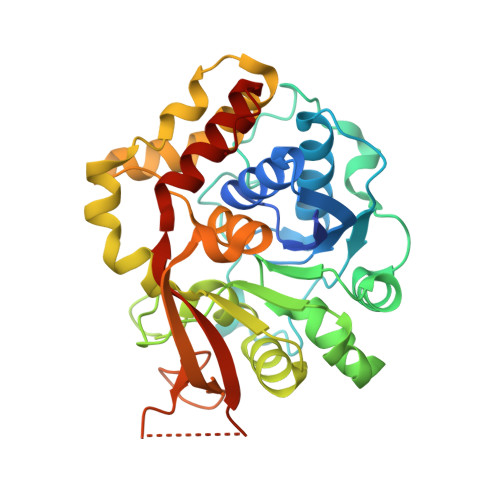
    5MJ7

  • PubMed Abstract: 

    Nucleoside hydrolases (NHs) catalyze the hydrolysis of the N-glycoside bond in ribonucleosides and are found in all three domains of life. Although in parasitic protozoa a role in purine salvage has been well established, their precise function in bacteria and higher eukaryotes is still largely unknown. NHs have been classified into three homology groups based on the conservation of active site residues. While many structures are available of representatives of group I and II, structural information for group III NHs is lacking. Here, we report the first crystal structure of a purine-specific nucleoside hydrolase belonging to homology group III from the nematode Caenorhabditis elegans (CeNH) to 1.65Å resolution. In contrast to dimeric purine-specific NHs from group II, CeNH is a homotetramer. A cysteine residue that characterizes group III NHs (Cys253) structurally aligns with the catalytic histidine and tryptophan residues of group I and group II enzymes, respectively. Moreover, a second cysteine (Cys42) points into the active site of CeNH. Substrate docking shows that both cysteine residues are appropriately positioned to interact with the purine ring. Site-directed mutagenesis and kinetic analysis proposes a catalytic role for both cysteines residues, with Cys253 playing the most prominent role in leaving group activation.


  • Organizational Affiliation

    Structural Biology Brussels, Vrije Universiteit Brussel (VUB), Pleinlaan 2, Brussels, 1050, Belgium.


Macromolecules
Find similar proteins by:  (by identity cutoff)  |  3D Structure
Entity ID: 1
MoleculeChains Sequence LengthOrganismDetailsImage
Uncharacterized protein
A, B
349Caenorhabditis elegansMutation(s): 0 
Gene Names: CELE_Y43F8C.13Y43F8C.13
UniProt
Find proteins for Q9XWN7 (Caenorhabditis elegans)
Explore Q9XWN7 
Go to UniProtKB:  Q9XWN7
Entity Groups  
Sequence Clusters30% Identity50% Identity70% Identity90% Identity95% Identity100% Identity
UniProt GroupQ9XWN7
Sequence Annotations
Expand
  • Reference Sequence
Experimental Data & Validation

Experimental Data

  • Method: X-RAY DIFFRACTION
  • Resolution: 1.65 Å
  • R-Value Free: 0.154 
  • R-Value Work: 0.137 
  • R-Value Observed: 0.138 
  • Space Group: P 41 21 2
Unit Cell:
Length ( Å )Angle ( ˚ )
a = 84.578α = 90
b = 84.578β = 90
c = 260.911γ = 90
Software Package:
Software NamePurpose
REFMACrefinement
DENZOdata reduction
SCALEPACKdata scaling
PHASERphasing

Structure Validation

View Full Validation Report


Entry History & Funding Information

Deposition Data


Funding OrganizationLocationGrant Number
Fonds voor Wetenschappelijk OnderzoekBelgium--
BioStruct-XBelgium--
Strategic Research Program- VUBBelgiumSRP34

Revision History  (Full details and data files)

  • Version 1.0: 2017-03-08
    Type: Initial release
  • Version 1.1: 2017-05-03
    Changes: Database references
  • Version 1.2: 2019-07-17
    Changes: Data collection
  • Version 1.3: 2024-01-17
    Changes: Advisory, Data collection, Database references, Derived calculations, Refinement description