5M99

Functional Characterization and Crystal Structure of Thermostable Amylase from Thermotoga petrophila, reveals High Thermostability and an Archaic form of Dimerization


Experimental Data Snapshot

  • Method: X-RAY DIFFRACTION
  • Resolution: 1.96 Å
  • R-Value Free: 0.184 
  • R-Value Work: 0.152 
  • R-Value Observed: 0.152 

wwPDB Validation   3D Report Full Report


This is version 1.1 of the entry. See complete history


Literature

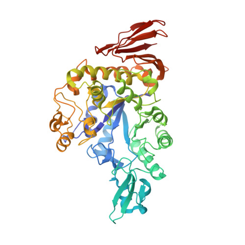
Functional characterization and crystal structure of thermostable amylase from Thermotoga petrophila, reveals high thermostability and an unusual form of dimerization.

Hameed, U.Price, I.Ke, A.Wilson, D.B.Mirza, O.

(2017) Biochim Biophys Acta 1865: 1237-1245

  • DOI: https://doi.org/10.1016/j.bbapap.2017.06.015
  • Primary Citation of Related Structures:  
    5M99

  • PubMed Abstract: 

    Thermostable α-amylases have many industrial applications and are therefore continuously explored from novel sources. We present the characterization of a novel putative α-amylase gene product (Tp-AmyS) cloned from Thermotoga petrophila. The purified recombinant enzyme is highly thermostable and able to hydrolyze starch into dextrin between 90 and 100°C, with optimum activity at 98°C and pH8.5. The activity increased in the presence of Rb 1+ , K 1+ and Ca 2+ ions, whereas other ions inhibited activity. The crystal structure of Tp-AmyS at 1.7Å resolution showed common features of the GH-13 family, however was apparently found to be a dimer. Several residues from one monomer interacted with a docked acarbose, an inhibitor of Tp-AmyS, in the other monomer, suggesting catalytic cooperativity within the dimer. The most striking feature of the dimer was that it resembled the dimerization of salivary amylase from a previous crystal structure, and thus could be a functional feature of some amylases.


  • Organizational Affiliation

    Institute of Industrial Biotechnology, Government College University, Lahore, 54000, Pakistan.


Macromolecules
Find similar proteins by:  (by identity cutoff)  |  3D Structure
Entity ID: 1
MoleculeChains Sequence LengthOrganismDetailsImage
Alpha-amylase506Thermotoga petrophila RKU-1Mutation(s): 0 
EC: 3.2.1.1
UniProt
Find proteins for A0A059TXD8 (Thermotoga petrophila (strain ATCC BAA-488 / DSM 13995 / JCM 10881 / RKU-1))
Explore A0A059TXD8 
Go to UniProtKB:  A0A059TXD8
Entity Groups  
Sequence Clusters30% Identity50% Identity70% Identity90% Identity95% Identity100% Identity
UniProt GroupA0A059TXD8
Sequence Annotations
Expand
  • Reference Sequence
Find similar proteins by:  (by identity cutoff)  |  3D Structure
Entity ID: 2
MoleculeChains Sequence LengthOrganismDetailsImage
Alpha-amylase505Thermotoga petrophila RKU-1Mutation(s): 0 
EC: 3.2.1.1
UniProt
Find proteins for A0A059TXD8 (Thermotoga petrophila (strain ATCC BAA-488 / DSM 13995 / JCM 10881 / RKU-1))
Explore A0A059TXD8 
Go to UniProtKB:  A0A059TXD8
Entity Groups  
Sequence Clusters30% Identity50% Identity70% Identity90% Identity95% Identity100% Identity
UniProt GroupA0A059TXD8
Sequence Annotations
Expand
  • Reference Sequence
Small Molecules
Ligands 5 Unique
IDChains Name / Formula / InChI Key2D Diagram3D Interactions
TRS
Query on TRS

Download Ideal Coordinates CCD File 
O [auth A]2-AMINO-2-HYDROXYMETHYL-PROPANE-1,3-DIOL
C4 H12 N O3
LENZDBCJOHFCAS-UHFFFAOYSA-O
EDO
Query on EDO

Download Ideal Coordinates CCD File 
AA [auth B]
BA [auth B]
CA [auth B]
DA [auth B]
EA [auth B]
AA [auth B],
BA [auth B],
CA [auth B],
DA [auth B],
EA [auth B],
I [auth A],
J [auth A],
K [auth A],
L [auth A],
M [auth A],
N [auth A],
V [auth B],
W [auth B],
X [auth B],
Y [auth B],
Z [auth B]
1,2-ETHANEDIOL
C2 H6 O2
LYCAIKOWRPUZTN-UHFFFAOYSA-N
CA
Query on CA

Download Ideal Coordinates CCD File 
H [auth A],
U [auth B]
CALCIUM ION
Ca
BHPQYMZQTOCNFJ-UHFFFAOYSA-N
K
Query on K

Download Ideal Coordinates CCD File 
G [auth A],
T [auth B]
POTASSIUM ION
K
NPYPAHLBTDXSSS-UHFFFAOYSA-N
NA
Query on NA

Download Ideal Coordinates CCD File 
C [auth A]
D [auth A]
E [auth A]
F [auth A]
P [auth B]
C [auth A],
D [auth A],
E [auth A],
F [auth A],
P [auth B],
Q [auth B],
R [auth B],
S [auth B]
SODIUM ION
Na
FKNQFGJONOIPTF-UHFFFAOYSA-N
Experimental Data & Validation

Experimental Data

  • Method: X-RAY DIFFRACTION
  • Resolution: 1.96 Å
  • R-Value Free: 0.184 
  • R-Value Work: 0.152 
  • R-Value Observed: 0.152 
  • Space Group: P 65
Unit Cell:
Length ( Å )Angle ( ˚ )
a = 153.237α = 90
b = 153.237β = 90
c = 143.056γ = 120
Software Package:
Software NamePurpose
PHENIXrefinement
XDSdata reduction
XDSdata scaling
PHASERphasing

Structure Validation

View Full Validation Report


Entry History 

Deposition Data

Revision History  (Full details and data files)

  • Version 1.0: 2017-07-19
    Type: Initial release
  • Version 1.1: 2017-08-09
    Changes: Database references