5M6S

folding intermediate of spectrin R16


Experimental Data Snapshot

  • Method: ELECTRON MICROSCOPY
  • Resolution: 4.80 Å
  • Aggregation State: PARTICLE 
  • Reconstruction Method: SINGLE PARTICLE 

wwPDB Validation   3D Report Full Report


This is version 1.4 of the entry. See complete history


Literature

Cotranslational folding of spectrin domains via partially structured states.

Nilsson, O.B.Nickson, A.A.Hollins, J.J.Wickles, S.Steward, A.Beckmann, R.von Heijne, G.Clarke, J.

(2017) Nat Struct Mol Biol 24: 221-225

  • DOI: https://doi.org/10.1038/nsmb.3355
  • Primary Citation of Related Structures:  
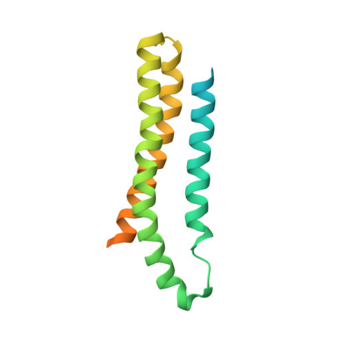
    5M6S

  • PubMed Abstract: 

    How do the key features of protein folding, elucidated from studies on native, isolated proteins, manifest in cotranslational folding on the ribosome? Using a well-characterized family of homologous α-helical proteins with a range of biophysical properties, we show that spectrin domains can fold vectorially on the ribosome and may do so via a pathway different from that of the isolated domain. We use cryo-EM to reveal a folded or partially folded structure, formed in the vestibule of the ribosome. Our results reveal that it is not possible to predict which domains will fold within the ribosome on the basis of the folding behavior of isolated domains; instead, we propose that a complex balance of the rate of folding, the rate of translation and the lifetime of folded or partly folded states will determine whether folding occurs cotranslationally on actively translating ribosomes.


  • Organizational Affiliation

    Department of Biochemistry and Biophysics Stockholm University, Stockholm, Sweden.


Macromolecules
Find similar proteins by:  (by identity cutoff)  |  3D Structure
Entity ID: 1
MoleculeChains Sequence LengthOrganismDetailsImage
spectrin169Escherichia coliMutation(s): 0 
UniProt
Find proteins for P0AD89 (Escherichia coli (strain K12))
Explore P0AD89 
Go to UniProtKB:  P0AD89
Find proteins for P07751 (Gallus gallus)
Explore P07751 
Go to UniProtKB:  P07751
Entity Groups  
Sequence Clusters30% Identity50% Identity70% Identity90% Identity95% Identity100% Identity
UniProt GroupsP07751P0AD89
Sequence Annotations
Expand
  • Reference Sequence
Experimental Data & Validation

Experimental Data

  • Method: ELECTRON MICROSCOPY
  • Resolution: 4.80 Å
  • Aggregation State: PARTICLE 
  • Reconstruction Method: SINGLE PARTICLE 
EM Software:
TaskSoftware PackageVersion
RECONSTRUCTIONSPIDER

Structure Validation

View Full Validation Report


Entry History 

Deposition Data

Revision History  (Full details and data files)

  • Version 1.0: 2017-01-11
    Type: Initial release
  • Version 1.1: 2017-01-25
    Changes: Database references
  • Version 1.2: 2017-02-01
    Changes: Database references
  • Version 1.3: 2017-03-15
    Changes: Database references
  • Version 1.4: 2017-08-02
    Changes: Data collection