5M1E

Crystal structure of N-terminally tagged UbiD from E. coli reconstituted with prFMN cofactor


Experimental Data Snapshot

  • Method: X-RAY DIFFRACTION
  • Resolution: 2.62 Å
  • R-Value Free: 0.212 
  • R-Value Work: 0.175 
  • R-Value Observed: 0.177 

wwPDB Validation   3D Report Full Report


Ligand Structure Quality Assessment 


This is version 1.3 of the entry. See complete history


Literature

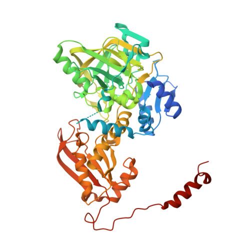
Oxidative Maturation and Structural Characterization of Prenylated FMN Binding by UbiD, a Decarboxylase Involved in Bacterial Ubiquinone Biosynthesis.

Marshall, S.A.Fisher, K.Ni Cheallaigh, A.White, M.D.Payne, K.A.Parker, D.A.Rigby, S.E.Leys, D.

(2017) J Biol Chem 292: 4623-4637

  • DOI: https://doi.org/10.1074/jbc.M116.762732
  • Primary Citation of Related Structures:  
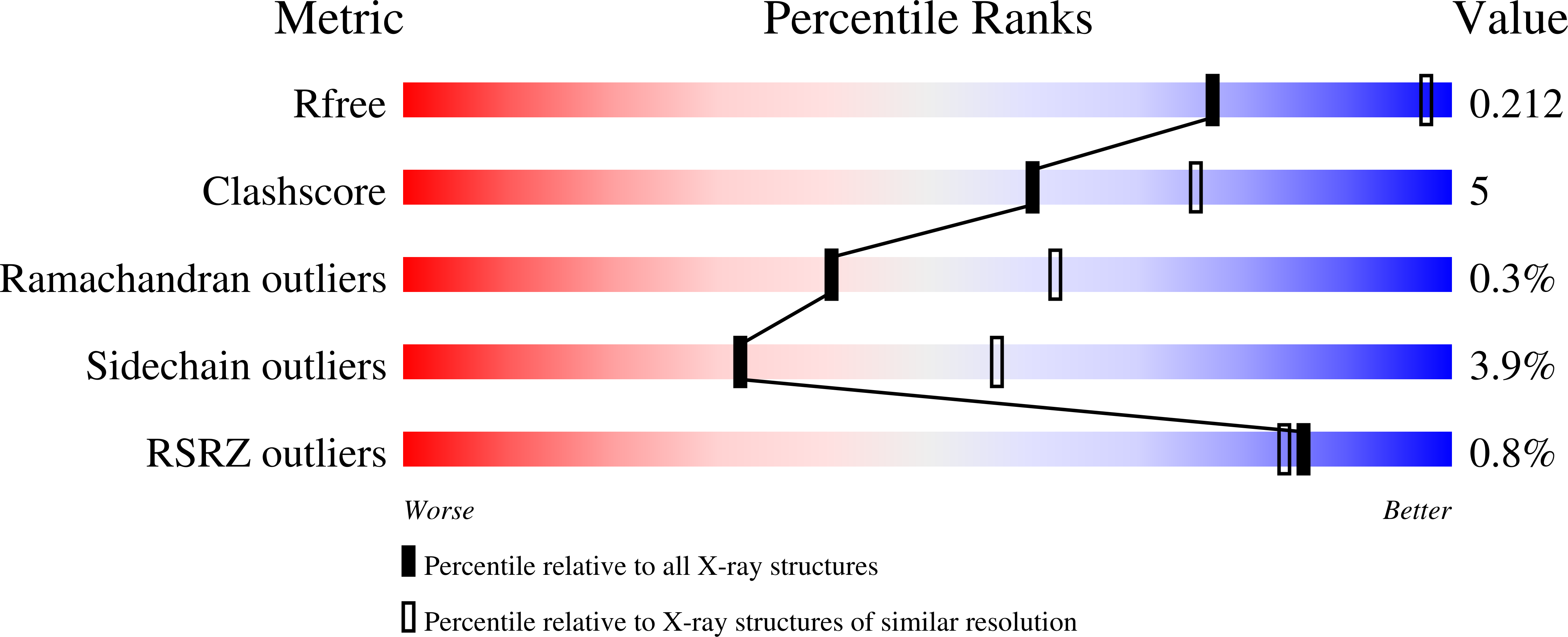
    5M1B, 5M1C, 5M1D, 5M1E

  • PubMed Abstract: 

    The activity of the reversible decarboxylase enzyme Fdc1 is dependent on prenylated FMN (prFMN), a recently discovered cofactor. The oxidized prFMN supports a 1,3-dipolar cycloaddition mechanism that underpins reversible decarboxylation. Fdc1 is a distinct member of the UbiD family of enzymes, with the canonical UbiD catalyzing the (de)carboxylation of para -hydroxybenzoic acid-type substrates. Here we show that the Escherichia coli UbiD enzyme, which is implicated in ubiquinone biosynthesis, cannot be isolated in an active holoenzyme form despite the fact active holoFdc1 is readily obtained. Formation of holoUbiD requires reconstitution in vitro of the apoUbiD with reduced prFMN. Furthermore, although the Fdc1 apoenzyme can be readily reconstituted and activated, in vitro oxidation to the mature prFMN cofactor stalls at formation of a radical prFMN species in holoUbiD. Further oxidative maturation in vitro occurs only at alkaline pH, suggesting a proton-coupled electron transfer precedes formation of the fully oxidized prFMN. Crystal structures of holoUbiD reveal a relatively open active site potentially occluded from solvent through domain motion. The presence of a prFMN sulfite-adduct in one of the UbiD crystal structures confirms oxidative maturation does occur at ambient pH on a slow time scale. Activity could not be detected for a range of putative para -hydroxybenzoic acid substrates tested. However, the lack of an obvious hydrophobic binding pocket for the octaprenyl tail of the proposed ubiquinone precursor substrate does suggest UbiD might act on a non-prenylated precursor. Our data reveals an unexpected variation occurs in domain mobility, prFMN binding, and maturation by the UbiD enzyme family.


  • Organizational Affiliation

    From the Manchester Institute of Biotechnology, University of Manchester, 131 Princess Street Manchester, M1 7DN, United Kingdom and.


Macromolecules
Find similar proteins by:  (by identity cutoff)  |  3D Structure
Entity ID: 1
MoleculeChains Sequence LengthOrganismDetailsImage
3-octaprenyl-4-hydroxybenzoate carboxy-lyase
A, B, C
517Escherichia coli CFT073Mutation(s): 0 
Gene Names: ubiDc4790
EC: 4.1.1.98
UniProt
Find proteins for P0AAB5 (Escherichia coli O6:H1 (strain CFT073 / ATCC 700928 / UPEC))
Explore P0AAB5 
Go to UniProtKB:  P0AAB5
Entity Groups  
Sequence Clusters30% Identity50% Identity70% Identity90% Identity95% Identity100% Identity
UniProt GroupP0AAB5
Sequence Annotations
Expand
  • Reference Sequence
Small Molecules
Ligands 3 Unique
IDChains Name / Formula / InChI Key2D Diagram3D Interactions
7D9
Query on 7D9

Download Ideal Coordinates CCD File 
E [auth A],
H [auth B],
K [auth C]
(16~{R})-11,12,14,14-tetramethyl-3,5-bis(oxidanylidene)-8-[(2~{S},3~{S},4~{R})-2,3,4-tris(oxidanyl)-5-phosphonooxy-pentyl]-1,4,6,8-tetrazatetracyclo[7.7.1.0^{2,7}.0^{13,17}]heptadeca-2(7),9(17),10,12-tetraene-16-sulfonic acid
C22 H31 N4 O12 P S
TZDQUUMSMXVHAK-LWGWVAHUSA-N
MN
Query on MN

Download Ideal Coordinates CCD File 
D [auth A],
G [auth B],
J [auth C]
MANGANESE (II) ION
Mn
WAEMQWOKJMHJLA-UHFFFAOYSA-N
NA
Query on NA

Download Ideal Coordinates CCD File 
F [auth A],
I [auth B],
L [auth C]
SODIUM ION
Na
FKNQFGJONOIPTF-UHFFFAOYSA-N
Experimental Data & Validation

Experimental Data

  • Method: X-RAY DIFFRACTION
  • Resolution: 2.62 Å
  • R-Value Free: 0.212 
  • R-Value Work: 0.175 
  • R-Value Observed: 0.177 
  • Space Group: P 43 21 2
Unit Cell:
Length ( Å )Angle ( ˚ )
a = 194.52α = 90
b = 194.52β = 90
c = 107.41γ = 90
Software Package:
Software NamePurpose
REFMACrefinement
xia2data reduction
xia2data scaling

Structure Validation

View Full Validation Report


Ligand Structure Quality Assessment 


Entry History 

Deposition Data

Revision History  (Full details and data files)

  • Version 1.0: 2017-01-11
    Type: Initial release
  • Version 1.1: 2017-01-18
    Changes: Database references
  • Version 1.2: 2017-03-29
    Changes: Database references
  • Version 1.3: 2024-01-17
    Changes: Data collection, Database references, Derived calculations, Refinement description