5LP8

Crystal structure of an asymmetric dimer of the ubiquitin ligase HUWE1


Experimental Data Snapshot

  • Method: X-RAY DIFFRACTION
  • Resolution: 2.70 Å
  • R-Value Free: 0.225 
  • R-Value Work: 0.194 
  • R-Value Observed: 0.195 

wwPDB Validation   3D Report Full Report


This is version 1.2 of the entry. See complete history


Literature

A conformational switch regulates the ubiquitin ligase HUWE1.

Sander, B.Xu, W.Eilers, M.Popov, N.Lorenz, S.

(2017) Elife 6

  • DOI: https://doi.org/10.7554/eLife.21036
  • Primary Citation of Related Structures:  
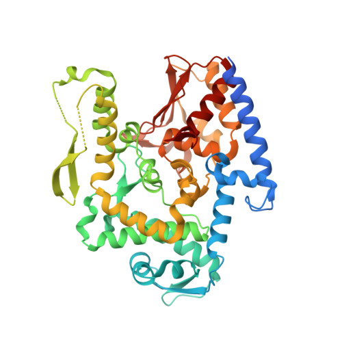
    5LP8

  • PubMed Abstract: 

    The human ubiquitin ligase HUWE1 has key roles in tumorigenesis, yet it is unkown how its activity is regulated. We present the crystal structure of a C-terminal part of HUWE1, including the catalytic domain, and reveal an asymmetric auto-inhibited dimer. We show that HUWE1 dimerizes in solution and self-associates in cells, and that both occurs through the crystallographic dimer interface. We demonstrate that HUWE1 is inhibited in cells and that it can be activated by disruption of the dimer interface. We identify a conserved segment in HUWE1 that counteracts dimer formation by associating with the dimerization region intramolecularly. Our studies reveal, intriguingly, that the tumor suppressor p14ARF binds to this segment and may thus shift the conformational equilibrium of HUWE1 toward the inactive state. We propose a model, in which the activity of HUWE1 underlies conformational control in response to physiological cues-a mechanism that may be exploited for cancer therapy.


  • Organizational Affiliation

    Rudolf Virchow Center for Experimental Biomedicine, University of Würzburg, Würzburg, Germany.


Macromolecules
Find similar proteins by:  (by identity cutoff)  |  3D Structure
Entity ID: 1
MoleculeChains Sequence LengthOrganismDetailsImage
E3 ubiquitin-protein ligase HUWE1A [auth B],
B [auth A]
450Homo sapiensMutation(s): 0 
Gene Names: HUWE1KIAA0312KIAA1578UREB1HSPC272
EC: 6.3.2
UniProt & NIH Common Fund Data Resources
Find proteins for Q7Z6Z7 (Homo sapiens)
Explore Q7Z6Z7 
Go to UniProtKB:  Q7Z6Z7
PHAROS:  Q7Z6Z7
GTEx:  ENSG00000086758 
Entity Groups  
Sequence Clusters30% Identity50% Identity70% Identity90% Identity95% Identity100% Identity
UniProt GroupQ7Z6Z7
Sequence Annotations
Expand
  • Reference Sequence
Experimental Data & Validation

Experimental Data

  • Method: X-RAY DIFFRACTION
  • Resolution: 2.70 Å
  • R-Value Free: 0.225 
  • R-Value Work: 0.194 
  • R-Value Observed: 0.195 
  • Space Group: P 63
Unit Cell:
Length ( Å )Angle ( ˚ )
a = 177.464α = 90
b = 177.464β = 90
c = 106.259γ = 120
Software Package:
Software NamePurpose
PHENIXrefinement
XDSdata reduction
Aimlessdata scaling
PHASERphasing

Structure Validation

View Full Validation Report


Entry History & Funding Information

Deposition Data


Funding OrganizationLocationGrant Number
German Research FoundationGermanyLO 2003/1-1

Revision History  (Full details and data files)

  • Version 1.0: 2017-02-22
    Type: Initial release
  • Version 1.1: 2017-04-12
    Changes: Data collection
  • Version 1.2: 2024-01-10
    Changes: Author supporting evidence, Data collection, Database references, Refinement description