5LHQ

The EGR-cmk active site inhibited catalytic domain of murine urokinase-type plasminogen activator in complex with the allosteric inhibitory nanobody Nb7


Experimental Data Snapshot

  • Method: X-RAY DIFFRACTION
  • Resolution: 2.60 Å
  • R-Value Free: 0.217 
  • R-Value Work: 0.177 
  • R-Value Observed: 0.179 

wwPDB Validation   3D Report Full Report


Ligand Structure Quality Assessment 


This is version 1.2 of the entry. See complete history


Literature

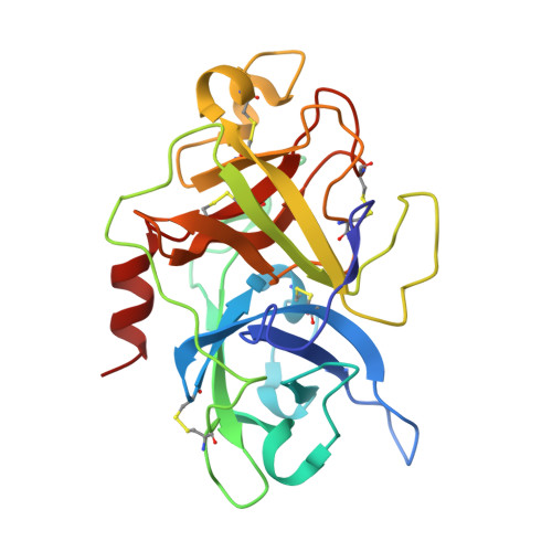
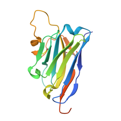
Discovery of a novel conformational equilibrium in urokinase-type plasminogen activator.

Kromann-Hansen, T.Louise Lange, E.Peter Srensen, H.Hassanzadeh-Ghassabeh, G.Huang, M.Jensen, J.K.Muyldermans, S.Declerck, P.J.Komives, E.A.Andreasen, P.A.

(2017) Sci Rep 7: 3385-3385

  • DOI: https://doi.org/10.1038/s41598-017-03457-7
  • Primary Citation of Related Structures:  
    5LHN, 5LHP, 5LHQ, 5LHR, 5LHS

  • PubMed Abstract: 

    Although trypsin-like serine proteases have flexible surface-exposed loops and are known to adopt higher and lower activity conformations, structural determinants for the different conformations have remained largely obscure. The trypsin-like serine protease, urokinase-type plasminogen activator (uPA), is central in tissue remodeling processes and also strongly implicated in tumor metastasis. We solved five X-ray crystal structures of murine uPA (muPA) in the absence and presence of allosteric molecules and/or substrate-like molecules. The structure of unbound muPA revealed an unsuspected non-chymotrypsin-like protease conformation in which two β-strands in the core of the protease domain undergoes a major antiparallel-to-parallel conformational transition. We next isolated two anti-muPA nanobodies; an active-site binding nanobody and an allosteric nanobody. Crystal structures of the muPA:nanobody complexes and hydrogen-deuterium exchange mass spectrometry revealed molecular insights about molecular factors controlling the antiparallel-to-parallel equilibrium in muPA. Together with muPA activity assays, the data provide valuable insights into regulatory mechanisms and conformational flexibility of uPA and trypsin-like serine proteases in general.


  • Organizational Affiliation

    From the Department of Chemistry and Biochemistry, University of California at San Diego, La Jolla, California, United States. tkromanntofting@ucsd.edu.


Macromolecules
Find similar proteins by:  (by identity cutoff)  |  3D Structure
Entity ID: 1
MoleculeChains Sequence LengthOrganismDetailsImage
Urokinase-type plasminogen activator247Mus musculusMutation(s): 1 
Gene Names: Plau
EC: 3.4.21.73
UniProt & NIH Common Fund Data Resources
Find proteins for P06869 (Mus musculus)
Explore P06869 
Go to UniProtKB:  P06869
IMPC:  MGI:97611
Entity Groups  
Sequence Clusters30% Identity50% Identity70% Identity90% Identity95% Identity100% Identity
UniProt GroupP06869
Sequence Annotations
Expand
  • Reference Sequence
Find similar proteins by:  (by identity cutoff)  |  3D Structure
Entity ID: 2
MoleculeChains Sequence LengthOrganismDetailsImage
Camelid-Derived Antibody Fragment Nb7152Vicugna pacosMutation(s): 0 
Entity Groups  
Sequence Clusters30% Identity50% Identity70% Identity90% Identity95% Identity100% Identity
Sequence Annotations
Expand
  • Reference Sequence
Small Molecules
Ligands 3 Unique
IDChains Name / Formula / InChI Key2D Diagram3D Interactions
0GJ
Query on 0GJ

Download Ideal Coordinates CCD File 
C [auth A]L-alpha-glutamyl-N-{(1S)-4-{[amino(iminio)methyl]amino}-1-[(1S)-2-chloro-1-hydroxyethyl]butyl}glycinamide
C14 H28 Cl N6 O5
XELWNHKFCNMWQO-LPEHRKFASA-O
SO4
Query on SO4

Download Ideal Coordinates CCD File 
D [auth A]
E [auth A]
F [auth A]
G [auth A]
H [auth A]
D [auth A],
E [auth A],
F [auth A],
G [auth A],
H [auth A],
I [auth A],
L [auth B],
M [auth B],
N [auth B],
O [auth B],
P [auth B]
SULFATE ION
O4 S
QAOWNCQODCNURD-UHFFFAOYSA-L
EDO
Query on EDO

Download Ideal Coordinates CCD File 
J [auth A],
K [auth A]
1,2-ETHANEDIOL
C2 H6 O2
LYCAIKOWRPUZTN-UHFFFAOYSA-N
Biologically Interesting Molecules (External Reference) 1 Unique
Experimental Data & Validation

Experimental Data

  • Method: X-RAY DIFFRACTION
  • Resolution: 2.60 Å
  • R-Value Free: 0.217 
  • R-Value Work: 0.177 
  • R-Value Observed: 0.179 
  • Space Group: P 31 2 1
Unit Cell:
Length ( Å )Angle ( ˚ )
a = 94.25α = 90
b = 94.25β = 90
c = 122.99γ = 120
Software Package:
Software NamePurpose
PHENIXrefinement
XDSdata reduction
XSCALEdata scaling
PHASERphasing

Structure Validation

View Full Validation Report


Ligand Structure Quality Assessment 


Entry History & Funding Information

Deposition Data


Funding OrganizationLocationGrant Number
Danish National Research FoundationDenmark26-331-6
Natural Science Foundation of ChinaChina31161130356, 31170707, 31370737
Lundbeck FoundationDenmarkR83-A7826
Carlsberg FoundationDenmarkCF15-0814

Revision History  (Full details and data files)

  • Version 1.0: 2017-06-28
    Type: Initial release
  • Version 1.1: 2018-06-20
    Changes: Data collection, Structure summary
  • Version 1.2: 2024-01-10
    Changes: Data collection, Database references, Refinement description