Structure function studies of R. palustris RubisCO.
Arbing, M.A., North, J.A., Satagopan, S., Varaljay, V.A., Shin, A., Tabita, F.R.To be published.
Experimental Data Snapshot
Entity ID: 1 | |||||
|---|---|---|---|---|---|
| Molecule | Chains | Sequence Length | Organism | Details | Image |
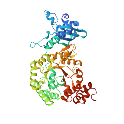
| Ribulose bisphosphate carboxylase | 481 | Rhodopseudomonas palustris | Mutation(s): 1 Gene Names: cbbM, RPA4641 EC: 4.1.1.39 |  | |
UniProt | |||||
Find proteins for Q6N0W9 (Rhodopseudomonas palustris (strain ATCC BAA-98 / CGA009)) Explore Q6N0W9 Go to UniProtKB: Q6N0W9 | |||||
Entity Groups | |||||
| Sequence Clusters | 30% Identity50% Identity70% Identity90% Identity95% Identity100% Identity | ||||
| UniProt Group | Q6N0W9 | ||||
Sequence AnnotationsExpand | |||||
| |||||
| Ligands 3 Unique | |||||
|---|---|---|---|---|---|
| ID | Chains | Name / Formula / InChI Key | 2D Diagram | 3D Interactions | |
| CAP Query on CAP | BA [auth F] EA [auth G] HA [auth H] KA [auth I] M [auth A] | 2-CARBOXYARABINITOL-1,5-DIPHOSPHATE C6 H14 O13 P2 ITHCSGCUQDMYAI-ZMIZWQJLSA-N |  | ||
| CO3 Query on CO3 | AA [auth E] DA [auth F] GA [auth G] JA [auth H] MA [auth I] | CARBONATE ION C O3 BVKZGUZCCUSVTD-UHFFFAOYSA-L |  | ||
| MG Query on MG | CA [auth F] FA [auth G] IA [auth H] LA [auth I] N [auth A] | MAGNESIUM ION Mg JLVVSXFLKOJNIY-UHFFFAOYSA-N |  | ||
| Length ( Å ) | Angle ( ˚ ) |
|---|---|
| a = 75.46 | α = 89.96 |
| b = 110.62 | β = 101.77 |
| c = 166.85 | γ = 104.88 |
| Software Name | Purpose |
|---|---|
| XDS | data reduction |
| XSCALE | data scaling |
| PHASER | phasing |
| PHENIX | refinement |
| PDB_EXTRACT | data extraction |
| Funding Organization | Location | Grant Number |
|---|---|---|
| National Institutes of Health/National Institute of General Medical Sciences (NIH/NIGMS) | United States | GM095742 |
| Department of Energy (DOE, United States) | United States | DE-FC02-02ER63421 |