5KNC

Crystal structure of the 3 ADP-bound V1 complex


Experimental Data Snapshot

  • Method: X-RAY DIFFRACTION
  • Resolution: 3.02 Å
  • R-Value Free: 0.253 
  • R-Value Work: 0.214 
  • R-Value Observed: 0.216 

wwPDB Validation   3D Report Full Report


Ligand Structure Quality Assessment 


This is version 1.3 of the entry. See complete history


Literature

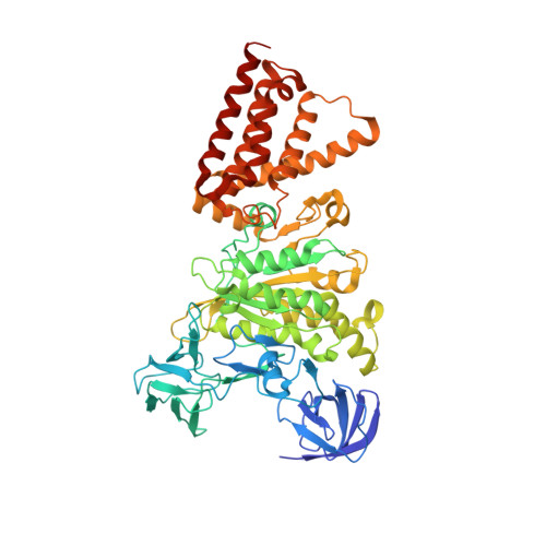
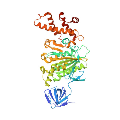
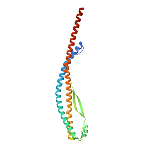
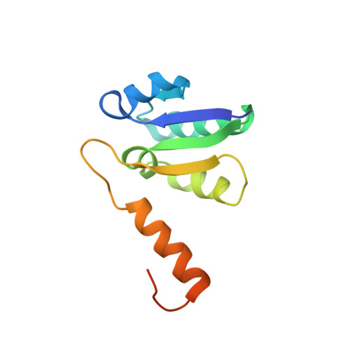
Crystal structures of the ATP-binding and ADP-release dwells of the V1 rotary motor

Suzuki, K.Mizutani, K.Maruyama, S.Shimono, K.Imai, F.L.Muneyuki, E.Kakinuma, Y.Ishizuka-Katsura, Y.Shirouzu, M.Yokoyama, S.Yamato, I.Murata, T.

(2016) Nat Commun 7: 13235-13235

  • DOI: https://doi.org/10.1038/ncomms13235
  • Primary Citation of Related Structures:  
    5KNB, 5KNC, 5KND

  • PubMed Abstract: 

    V 1 -ATPases are highly conserved ATP-driven rotary molecular motors found in various membrane systems. We recently reported the crystal structures for the Enterococcus hirae A 3 B 3 DF (V 1 ) complex, corresponding to the catalytic dwell state waiting for ATP hydrolysis. Here we present the crystal structures for two other dwell states obtained by soaking nucleotide-free V 1 crystals in ADP. In the presence of 20 μM ADP, two ADP molecules bind to two of three binding sites and cooperatively induce conformational changes of the third site to an ATP-binding mode, corresponding to the ATP-binding dwell. In the presence of 2 mM ADP, all nucleotide-binding sites are occupied by ADP to induce conformational changes corresponding to the ADP-release dwell. Based on these and previous findings, we propose a V 1 -ATPase rotational mechanism model.


  • Organizational Affiliation

    Department of Chemistry, Graduate School of Science, Chiba University, 1-33 Yayoi-cho, Inage, Chiba 263-8522, Japan.


Macromolecules
Find similar proteins by:  (by identity cutoff)  |  3D Structure
Entity ID: 1
MoleculeChains Sequence LengthOrganismDetailsImage
V-type sodium ATPase catalytic subunit A
A, B, C
600Enterococcus hirae ATCC 9790Mutation(s): 0 
Gene Names: ntpA
EC: 3.6.3.15
UniProt
Find proteins for Q08636 (Enterococcus hirae (strain ATCC 9790 / DSM 20160 / JCM 8729 / LMG 6399 / NBRC 3181 / NCIMB 6459 / NCDO 1258 / NCTC 12367 / WDCM 00089 / R))
Explore Q08636 
Go to UniProtKB:  Q08636
Entity Groups  
Sequence Clusters30% Identity50% Identity70% Identity90% Identity95% Identity100% Identity
UniProt GroupQ08636
Sequence Annotations
Expand
  • Reference Sequence
Find similar proteins by:  (by identity cutoff)  |  3D Structure
Entity ID: 2
MoleculeChains Sequence LengthOrganismDetailsImage
V-type sodium ATPase subunit B
D, E, F
465Enterococcus hirae ATCC 9790Mutation(s): 0 
Gene Names: ntpB
UniProt
Find proteins for Q08637 (Enterococcus hirae (strain ATCC 9790 / DSM 20160 / JCM 8729 / LMG 6399 / NBRC 3181 / NCIMB 6459 / NCDO 1258 / NCTC 12367 / WDCM 00089 / R))
Explore Q08637 
Go to UniProtKB:  Q08637
Entity Groups  
Sequence Clusters30% Identity50% Identity70% Identity90% Identity95% Identity100% Identity
UniProt GroupQ08637
Sequence Annotations
Expand
  • Reference Sequence
Find similar proteins by:  (by identity cutoff)  |  3D Structure
Entity ID: 3
MoleculeChains Sequence LengthOrganismDetailsImage
V-type sodium ATPase subunit D217Enterococcus hirae ATCC 9790Mutation(s): 0 
Gene Names: ntpD
UniProt
Find proteins for P43435 (Enterococcus hirae (strain ATCC 9790 / DSM 20160 / JCM 8729 / LMG 6399 / NBRC 3181 / NCIMB 6459 / NCDO 1258 / NCTC 12367 / WDCM 00089 / R))
Explore P43435 
Go to UniProtKB:  P43435
Entity Groups  
Sequence Clusters30% Identity50% Identity70% Identity90% Identity95% Identity100% Identity
UniProt GroupP43435
Sequence Annotations
Expand
  • Reference Sequence
Find similar proteins by:  (by identity cutoff)  |  3D Structure
Entity ID: 4
MoleculeChains Sequence LengthOrganismDetailsImage
V-type sodium ATPase subunit NtpG (F)115Enterococcus hirae ATCC 9790Mutation(s): 0 
Gene Names: ntpG
UniProt
Find proteins for P43455 (Enterococcus hirae (strain ATCC 9790 / DSM 20160 / JCM 8729 / LMG 6399 / NBRC 3181 / NCIMB 6459 / NCDO 1258 / NCTC 12367 / WDCM 00089 / R))
Explore P43455 
Go to UniProtKB:  P43455
Entity Groups  
Sequence Clusters30% Identity50% Identity70% Identity90% Identity95% Identity100% Identity
UniProt GroupP43455
Sequence Annotations
Expand
  • Reference Sequence
Small Molecules
Ligands 4 Unique
IDChains Name / Formula / InChI Key2D Diagram3D Interactions
ADP
Query on ADP

Download Ideal Coordinates CCD File 
K [auth A],
S [auth B],
Z [auth C]
ADENOSINE-5'-DIPHOSPHATE
C10 H15 N5 O10 P2
XTWYTFMLZFPYCI-KQYNXXCUSA-N
SO4
Query on SO4

Download Ideal Coordinates CCD File 
J [auth A]SULFATE ION
O4 S
QAOWNCQODCNURD-UHFFFAOYSA-L
GOL
Query on GOL

Download Ideal Coordinates CCD File 
AA [auth D]
BA [auth D]
CA [auth F]
L [auth A]
M [auth A]
AA [auth D],
BA [auth D],
CA [auth F],
L [auth A],
M [auth A],
N [auth A],
O [auth A],
P [auth A],
Q [auth A],
T [auth B],
U [auth B],
V [auth B],
W [auth B],
X [auth B]
GLYCEROL
C3 H8 O3
PEDCQBHIVMGVHV-UHFFFAOYSA-N
MG
Query on MG

Download Ideal Coordinates CCD File 
I [auth A],
R [auth B],
Y [auth C]
MAGNESIUM ION
Mg
JLVVSXFLKOJNIY-UHFFFAOYSA-N
Experimental Data & Validation

Experimental Data

  • Method: X-RAY DIFFRACTION
  • Resolution: 3.02 Å
  • R-Value Free: 0.253 
  • R-Value Work: 0.214 
  • R-Value Observed: 0.216 
  • Space Group: P 21 21 21
Unit Cell:
Length ( Å )Angle ( ˚ )
a = 121.68α = 90
b = 126.49β = 90
c = 225.26γ = 90
Software Package:
Software NamePurpose
PHENIXrefinement
XDSdata scaling
PHASERphasing
PDB_EXTRACTdata extraction
Cootmodel building

Structure Validation

View Full Validation Report


Ligand Structure Quality Assessment 


Entry History 

Revision History  (Full details and data files)

  • Version 1.0: 2016-11-02
    Type: Initial release
  • Version 1.1: 2016-11-16
    Changes: Database references
  • Version 1.2: 2020-02-19
    Changes: Data collection, Derived calculations
  • Version 1.3: 2023-11-08
    Changes: Data collection, Database references, Derived calculations, Refinement description