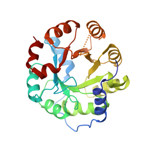
5K7J

Structure of designed zinc binding protein ZE2 bound to Zn2+


Experimental Data Snapshot

  • Method: X-RAY DIFFRACTION
  • Resolution: 1.39 Å
  • R-Value Free: 0.230 
  • R-Value Work: 0.196 
  • R-Value Observed: 0.198 

wwPDB Validation   3D Report Full Report


This is version 1.5 of the entry. See complete history


Literature

Probing the minimal determinants of zinc binding with computational protein design.

Guffy, S.L.Der, B.S.Kuhlman, B.

(2016) Protein Eng Des Sel 29: 327-338

  • DOI: https://doi.org/10.1093/protein/gzw026
  • Primary Citation of Related Structures:  
    5K7J, 5KAY

  • PubMed Abstract: 

    Structure-based protein design tests our understanding of the minimal determinants of protein structure and function. Previous studies have demonstrated that placing zinc binding amino acids (His, Glu, Asp or Cys) near each other in a folded protein in an arrangement predicted to be tetrahedral is often sufficient to achieve binding to zinc. However, few designs have been characterized with high-resolution structures. Here, we use X-ray crystallography, binding studies and mutation analysis to evaluate three alternative strategies for designing zinc binding sites with the molecular modeling program Rosetta. While several of the designs were observed to bind zinc, crystal structures of two designs reveal binding configurations that differ from the design model. In both cases, the modeling did not accurately capture the presence or absence of second-shell hydrogen bonds critical in determining binding-site structure. Efforts to more explicitly design second-shell hydrogen bonds were largely unsuccessful as evidenced by mutation analysis and low expression of proteins engineered with extensive primary and secondary networks. Our results suggest that improved methods for designing interaction networks will be needed for creating metal binding sites with high accuracy.


  • Organizational Affiliation

    Department of Biochemistry and Biophysics, University of North Carolina, Chapel Hill, NC 27599-7260, USA.


Macromolecules
Find similar proteins by:  (by identity cutoff)  |  3D Structure
Entity ID: 1
MoleculeChains Sequence LengthOrganismDetailsImage
Indole-3-glycerol phosphate synthase
A, B
249Saccharolobus solfataricus P2Mutation(s): 7 
Gene Names: trpCSSO0895
EC: 4.1.1.48
UniProt
Find proteins for Q06121 (Saccharolobus solfataricus (strain ATCC 35092 / DSM 1617 / JCM 11322 / P2))
Explore Q06121 
Go to UniProtKB:  Q06121
Entity Groups  
Sequence Clusters30% Identity50% Identity70% Identity90% Identity95% Identity100% Identity
UniProt GroupQ06121
Sequence Annotations
Expand
  • Reference Sequence
Experimental Data & Validation

Experimental Data

  • Method: X-RAY DIFFRACTION
  • Resolution: 1.39 Å
  • R-Value Free: 0.230 
  • R-Value Work: 0.196 
  • R-Value Observed: 0.198 
  • Space Group: P 1 21 1
Unit Cell:
Length ( Å )Angle ( ˚ )
a = 41.718α = 90
b = 79.029β = 95.7
c = 74.077γ = 90
Software Package:
Software NamePurpose
HKL-2000data collection
SCALEPACKdata scaling
PHASERphasing
REFMACrefinement
PDB_EXTRACTdata extraction
PHASERphasing
DENZOdata reduction

Structure Validation

View Full Validation Report


Entry History & Funding Information

Deposition Data


Funding OrganizationLocationGrant Number
National Institutes of Health/National Institute of General Medical Sciences (NIH/NIGMS)United StatesGM117968
National Science Foundation (NSF, United States)United StatesCBET-1403663

Revision History  (Full details and data files)

  • Version 1.0: 2016-08-03
    Type: Initial release
  • Version 1.1: 2016-08-10
    Changes: Database references
  • Version 1.2: 2017-09-06
    Changes: Author supporting evidence, Derived calculations
  • Version 1.3: 2017-11-01
    Changes: Author supporting evidence
  • Version 1.4: 2019-11-27
    Changes: Author supporting evidence
  • Version 1.5: 2023-09-27
    Changes: Data collection, Database references, Refinement description