5JST

MBP fused MDV1 coiled coil


Experimental Data Snapshot

  • Method: X-RAY DIFFRACTION
  • Resolution: 2.20 Å
  • R-Value Free: 0.274 
  • R-Value Work: 0.225 
  • R-Value Observed: 0.227 

wwPDB Validation   3D Report Full Report


This is version 2.1 of the entry. See complete history


Literature

ACCORD: an assessment tool to determine the orientation of homodimeric coiled-coils.

Kim, B.W.Jung, Y.O.Kim, M.K.Kwon, D.H.Park, S.H.Kim, J.H.Kuk, Y.B.Oh, S.J.Kim, L.Kim, B.H.Yang, W.S.Song, H.K.

(2017) Sci Rep 7: 43318-43318

  • DOI: https://doi.org/10.1038/srep43318
  • Primary Citation of Related Structures:  
    5JST

  • PubMed Abstract: 

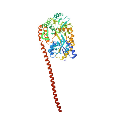
    The coiled-coil (CC) domain is a very important structural unit of proteins that plays critical roles in various biological functions. The major oligomeric state of CCs is a dimer, which can be either parallel or antiparallel. The orientation of each α-helix in a CC domain is critical for the molecular function of CC-containing proteins, but cannot be determined easily by sequence-based prediction. We developed a biochemical method for assessing differences between parallel and antiparallel CC homodimers and named it ACCORD (Assessment tool for homodimeric Coiled-Coil ORientation Decision). To validate this technique, we applied it to 15 different CC proteins with known structures, and the ACCORD results identified these proteins well, especially with long CCs. Furthermore, ACCORD was able to accurately determine the orientation of a CC domain of unknown directionality that was subsequently confirmed by X-ray crystallography and small angle X-ray scattering. Thus, ACCORD can be used as a tool to determine CC directionality to supplement the results of in silico prediction.


  • Organizational Affiliation

    Division of Life Sciences, Korea University, 145 Anam-ro, Seongbuk-gu, Seoul 02841, Korea.


Macromolecules
Find similar proteins by:  (by identity cutoff)  |  3D Structure
Entity ID: 1
MoleculeChains Sequence LengthOrganismDetailsImage
Maltose-binding periplasmic protein,Mitochondrial division protein 1
A, B
443Escherichia coli K-12Saccharomyces cerevisiae YJM789
This entity is chimeric
Mutation(s): 6 
Gene Names: malEb4034JW3994MDV1FIS2GAG3NET2SCY_3176
UniProt
Find proteins for P0AEX9 (Escherichia coli (strain K12))
Explore P0AEX9 
Go to UniProtKB:  P0AEX9
Find proteins for A6ZQL5 (Saccharomyces cerevisiae (strain YJM789))
Explore A6ZQL5 
Go to UniProtKB:  A6ZQL5
Entity Groups  
Sequence Clusters30% Identity50% Identity70% Identity90% Identity95% Identity100% Identity
UniProt GroupsA6ZQL5P0AEX9
Sequence Annotations
Expand
  • Reference Sequence
Oligosaccharides

Help

Entity ID: 2
MoleculeChains Length2D Diagram Glycosylation3D Interactions
alpha-D-glucopyranose-(1-4)-alpha-D-glucopyranose
C, D
2N/A
Glycosylation Resources
GlyTouCan:  G07411ON
GlyCosmos:  G07411ON
Biologically Interesting Molecules (External Reference) 1 Unique
Experimental Data & Validation

Experimental Data

  • Method: X-RAY DIFFRACTION
  • Resolution: 2.20 Å
  • R-Value Free: 0.274 
  • R-Value Work: 0.225 
  • R-Value Observed: 0.227 
  • Space Group: C 1 2 1
Unit Cell:
Length ( Å )Angle ( ˚ )
a = 128.515α = 90
b = 102.942β = 102.51
c = 79.267γ = 90
Software Package:
Software NamePurpose
PHENIXrefinement
HKL-2000data scaling
PDB_EXTRACTdata extraction
HKL-2000data reduction
PHENIXphasing

Structure Validation

View Full Validation Report


Entry History 

Deposition Data

Revision History  (Full details and data files)

  • Version 1.0: 2017-03-22
    Type: Initial release
  • Version 2.0: 2020-07-29
    Type: Remediation
    Reason: Carbohydrate remediation
    Changes: Advisory, Atomic model, Data collection, Derived calculations, Non-polymer description, Structure summary
  • Version 2.1: 2023-11-08
    Changes: Data collection, Database references, Refinement description, Structure summary