


The crystal structure of azoreductase from Yersinia pestis CO92 in its Apo form
Tan, K., Gu, M., Kwon, K., Anderson, W.F., Joachimiak, A.To be published.
Experimental Data Snapshot
wwPDB Validation 3D Report Full Report

Entity ID: 1 | |||||
|---|---|---|---|---|---|
| Molecule | Chains | Sequence Length | Organism | Details | Image |
| FMN-dependent NADH-azoreductase | 204 | Yersinia pestis CO92 | Mutation(s): 0 Gene Names: azoR, YPO2323, y2010, YP_2110 EC: 1.7 | 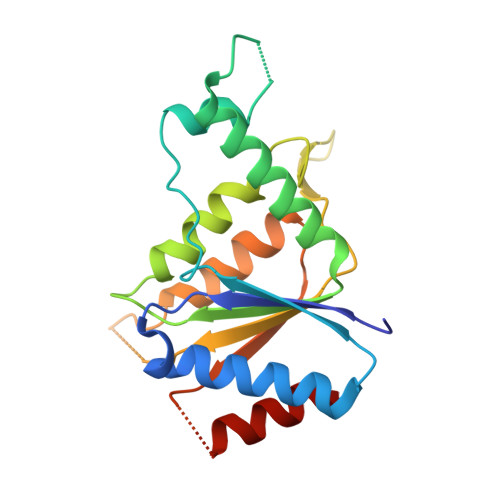 | |
UniProt | |||||
Find proteins for Q8ZE60 (Yersinia pestis) Explore Q8ZE60 Go to UniProtKB: Q8ZE60 | |||||
Entity Groups | |||||
| Sequence Clusters | 30% Identity50% Identity70% Identity90% Identity95% Identity100% Identity | ||||
| UniProt Group | Q8ZE60 | ||||
Sequence AnnotationsExpand | |||||
| |||||
| Ligands 1 Unique | |||||
|---|---|---|---|---|---|
| ID | Chains | Name / Formula / InChI Key | 2D Diagram | 3D Interactions | |
| GOL Query on GOL | C [auth A], D [auth B] | GLYCEROL C3 H8 O3 PEDCQBHIVMGVHV-UHFFFAOYSA-N |  | ||
| Length ( Å ) | Angle ( ˚ ) |
|---|---|
| a = 124.257 | α = 90 |
| b = 45.135 | β = 112.84 |
| c = 72.699 | γ = 90 |
| Software Name | Purpose |
|---|---|
| PHENIX | refinement |
| HKL-3000 | data reduction |
| HKL-3000 | data scaling |
| HKL-3000 | phasing |
| Funding Organization | Location | Grant Number |
|---|---|---|
| National Institutes of Health/National Institute Of Allergy and Infectious Diseases (NIH/NIAID) | United States | HHSN272201200026C |
RCSB PDB (citation) is hosted by
RCSB PDB is a member of the

