5JR7

Crystal structure of an ACRDYS heterodimer [RIa(92-365):C] of PKA


Experimental Data Snapshot

  • Method: X-RAY DIFFRACTION
  • Resolution: 3.56 Å
  • R-Value Free: 0.322 
  • R-Value Work: 0.266 
  • R-Value Observed: 0.269 

wwPDB Validation   3D Report Full Report


Ligand Structure Quality Assessment 


This is version 1.3 of the entry. See complete history


Literature

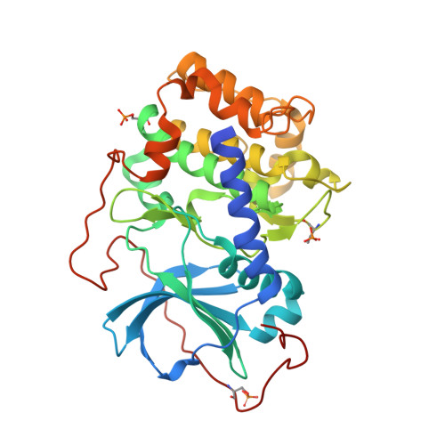
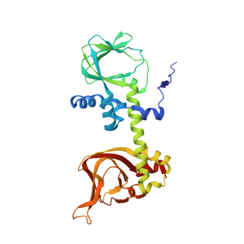
Structure of a PKA RI alpha Recurrent Acrodysostosis Mutant Explains Defective cAMP-Dependent Activation.

Bruystens, J.G.Wu, J.Fortezzo, A.Del Rio, J.Nielsen, C.Blumenthal, D.K.Rock, R.Stefan, E.Taylor, S.S.

(2016) J Mol Biol 428: 4890-4904

  • DOI: https://doi.org/10.1016/j.jmb.2016.10.033
  • Primary Citation of Related Structures:  
    5JR7

  • PubMed Abstract: 

    Most disease-related mutations that impair cAMP protein kinase A (PKA) signaling are present within the regulatory (R) PKA RI alpha-subunit (RIα). Although mutations in the PRKAR1A gene are linked to Carney complex (CNC) disease and, more recently, to acrodysostosis-1 (ACRDYS1), the two diseases show contrasting phenotypes. While CNC mutations cause increased PKA activity, ACRDYS1 mutations result in decreased PKA activity and cAMP resistant holoenzymes. Mapping the ACRDYS1 disease mutations reveals their localization to the second of two tandem cAMP-binding (CNB) domains (CNB-B), and here, we characterize a recurrent deletion mutant where the last 14 residues are missing. The crystal structure of a monomeric form of this mutant (RIα92-365) bound to the catalytic (C)-subunit reveals the dysfunctional regions of the RIα subunit. Beyond the missing residues, the entire capping motif is disordered (residues 357-379) and explains the disrupted cAMP binding. Moreover, the effects of the mutation extend far beyond the CNB-B domain and include the active site and N-lobe of the C-subunit, which is in a partially open conformation with the C-tail disordered. A key residue that contributes to this crosstalk, D267, is altered in our structure, and we confirmed its functional importance by mutagenesis. In particular, the D267 interaction with Arg241, a residue shown earlier to be important for allosteric regulation, is disrupted, thereby strengthening the interaction of D267 with the C-subunit residue Arg194 at the R:C interface. We see here how the switch between active (cAMP-bound) and inactive (holoenzyme) conformations is perturbed and how the dynamically controlled crosstalk between the helical domains of the two CNB domains is necessary for the functional regulation of PKA activity.


  • Organizational Affiliation

    Department of Chemistry and Biochemistry, University of California at San Diego, La Jolla, CA 92093, USA.


Macromolecules
Find similar proteins by:  (by identity cutoff)  |  3D Structure
Entity ID: 1
MoleculeChains Sequence LengthOrganismDetailsImage
cAMP-dependent protein kinase catalytic subunit alpha
A, C
350Mus musculusMutation(s): 0 
Gene Names: PrkacaPkaca
EC: 2.7.11.11
UniProt
Find proteins for P05132 (Mus musculus)
Explore P05132 
Go to UniProtKB:  P05132
Entity Groups  
Sequence Clusters30% Identity50% Identity70% Identity90% Identity95% Identity100% Identity
UniProt GroupP05132
Sequence Annotations
Expand
  • Reference Sequence
Find similar proteins by:  (by identity cutoff)  |  3D Structure
Entity ID: 2
MoleculeChains Sequence LengthOrganismDetailsImage
cAMP-dependent protein kinase type I-alpha regulatory subunit
B, D
275Bos taurusMutation(s): 0 
Gene Names: PRKAR1A
UniProt
Find proteins for P00514 (Bos taurus)
Explore P00514 
Go to UniProtKB:  P00514
Entity Groups  
Sequence Clusters30% Identity50% Identity70% Identity90% Identity95% Identity100% Identity
UniProt GroupP00514
Sequence Annotations
Expand
  • Reference Sequence
Small Molecules
Modified Residues  2 Unique
IDChains TypeFormula2D DiagramParent
SEP
Query on SEP
A, C
L-PEPTIDE LINKINGC3 H8 N O6 PSER
TPO
Query on TPO
A, C
L-PEPTIDE LINKINGC4 H10 N O6 PTHR
Experimental Data & Validation

Experimental Data

  • Method: X-RAY DIFFRACTION
  • Resolution: 3.56 Å
  • R-Value Free: 0.322 
  • R-Value Work: 0.266 
  • R-Value Observed: 0.269 
  • Space Group: P 1
Unit Cell:
Length ( Å )Angle ( ˚ )
a = 60.146α = 101.76
b = 66.79β = 89.37
c = 87.932γ = 105.27
Software Package:
Software NamePurpose
PHENIXrefinement
HKL-2000data reduction
SCALEPACKdata scaling
PHASERphasing

Structure Validation

View Full Validation Report


Ligand Structure Quality Assessment 


Entry History & Funding Information

Deposition Data


Funding OrganizationLocationGrant Number
National Institutes of Health/National Institute of General Medical Sciences (NIH/NIGMS)United StatesGM 034921

Revision History  (Full details and data files)

  • Version 1.0: 2017-03-15
    Type: Initial release
  • Version 1.1: 2017-09-27
    Changes: Author supporting evidence
  • Version 1.2: 2019-12-25
    Changes: Author supporting evidence
  • Version 1.3: 2023-09-27
    Changes: Advisory, Data collection, Database references, Refinement description