5JNW

Crystal structure of bovine low molecular weight protein tyrosine phosphatase (LMPTP) mutant (W49Y N50E) complexed with vanadate and uncompetitive inhibitor


Experimental Data Snapshot

  • Method: X-RAY DIFFRACTION
  • Resolution: 1.86 Å
  • R-Value Free: 0.209 
  • R-Value Work: 0.166 
  • R-Value Observed: 0.168 

wwPDB Validation   3D Report Full Report


Ligand Structure Quality Assessment 


This is version 1.5 of the entry. See complete history


Literature

Diabetes reversal by inhibition of the low-molecular-weight tyrosine phosphatase.

Stanford, S.M.Aleshin, A.E.Zhang, V.Ardecky, R.J.Hedrick, M.P.Zou, J.Ganji, S.R.Bliss, M.R.Yamamoto, F.Bobkov, A.A.Kiselar, J.Liu, Y.Cadwell, G.W.Khare, S.Yu, J.Barquilla, A.Chung, T.D.Y.Mustelin, T.Schenk, S.Bankston, L.A.Liddington, R.C.Pinkerton, A.B.Bottini, N.

(2017) Nat Chem Biol 13: 624-632

  • DOI: https://doi.org/10.1038/nchembio.2344
  • Primary Citation of Related Structures:  
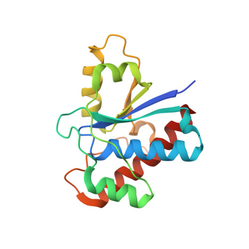
    5JNR, 5JNS, 5JNT, 5JNU, 5JNV, 5JNW

  • PubMed Abstract: 

    Obesity-associated insulin resistance plays a central role in type 2 diabetes. As such, tyrosine phosphatases that dephosphorylate the insulin receptor (IR) are potential therapeutic targets. The low-molecular-weight protein tyrosine phosphatase (LMPTP) is a proposed IR phosphatase, yet its role in insulin signaling in vivo has not been defined. Here we show that global and liver-specific LMPTP deletion protects mice from high-fat diet-induced diabetes without affecting body weight. To examine the role of the catalytic activity of LMPTP, we developed a small-molecule inhibitor with a novel uncompetitive mechanism, a unique binding site at the opening of the catalytic pocket, and an exquisite selectivity over other phosphatases. This inhibitor is orally bioavailable, and it increases liver IR phosphorylation in vivo and reverses high-fat diet-induced diabetes. Our findings suggest that LMPTP is a key promoter of insulin resistance and that LMPTP inhibitors would be beneficial for treating type 2 diabetes.


  • Organizational Affiliation

    Division of Cellular Biology, La Jolla Institute for Allergy and Immunology, La Jolla, California, USA.


Macromolecules
Find similar proteins by:  (by identity cutoff)  |  3D Structure
Entity ID: 1
MoleculeChains Sequence LengthOrganismDetailsImage
Low molecular weight phosphotyrosine protein phosphatase159Bos taurusMutation(s): 0 
Gene Names: ACP1
EC: 3.1.3.48
UniProt
Find proteins for P11064 (Bos taurus)
Explore P11064 
Go to UniProtKB:  P11064
Entity Groups  
Sequence Clusters30% Identity50% Identity70% Identity90% Identity95% Identity100% Identity
UniProt GroupP11064
Sequence Annotations
Expand
  • Reference Sequence
Small Molecules
Ligands 2 Unique
IDChains Name / Formula / InChI Key2D Diagram3D Interactions
6LJ
Query on 6LJ

Download Ideal Coordinates CCD File 
C [auth A]2-(4-{[3-(piperidin-1-yl)propyl]amino}quinolin-2-yl)benzonitrile
C24 H26 N4
PBIFICWHSUFSCR-UHFFFAOYSA-N
VO4
Query on VO4

Download Ideal Coordinates CCD File 
B [auth A]VANADATE ION
O4 V
LSGOVYNHVSXFFJ-UHFFFAOYSA-N
Binding Affinity Annotations 
IDSourceBinding Affinity
6LJ Binding MOAD:  5JNW IC50: 460 (nM) from 1 assay(s)
Experimental Data & Validation

Experimental Data

  • Method: X-RAY DIFFRACTION
  • Resolution: 1.86 Å
  • R-Value Free: 0.209 
  • R-Value Work: 0.166 
  • R-Value Observed: 0.168 
  • Space Group: C 2 2 21
Unit Cell:
Length ( Å )Angle ( ˚ )
a = 60.347α = 90
b = 125.493β = 90
c = 45.251γ = 90
Software Package:
Software NamePurpose
REFMACrefinement
Aimlessdata scaling
PDB_EXTRACTdata extraction
XDSdata reduction
PHASERphasing

Structure Validation

View Full Validation Report


Ligand Structure Quality Assessment 


Entry History & Funding Information

Deposition Data


Funding OrganizationLocationGrant Number
National Institutes of Health/National Institute of Diabetes and Digestive and Kidney Disease (NIH/NIDDK)United StatesR01DK106233

Revision History  (Full details and data files)

  • Version 1.0: 2017-03-29
    Type: Initial release
  • Version 1.1: 2017-04-12
    Changes: Database references
  • Version 1.2: 2017-05-24
    Changes: Database references
  • Version 1.3: 2017-09-20
    Changes: Author supporting evidence
  • Version 1.4: 2019-12-25
    Changes: Author supporting evidence, Derived calculations
  • Version 1.5: 2023-09-27
    Changes: Data collection, Database references, Refinement description