5J51

Structure of tetrameric jacalin complexed with Gal alpha-(1,4) Gal


Experimental Data Snapshot

  • Method: X-RAY DIFFRACTION
  • Resolution: 1.67 Å
  • R-Value Free: 0.213 
  • R-Value Work: 0.164 
  • R-Value Observed: 0.167 

wwPDB Validation   3D Report Full Report


This is version 2.1 of the entry. See complete history


Literature

Effect of linkage on the location of reducing and nonreducing sugars bound to jacalin.

Abhinav, K.V.Sharma, K.Surolia, A.Vijayan, M.

(2016) IUBMB Life 68: 971-979

  • DOI: https://doi.org/10.1002/iub.1572
  • Primary Citation of Related Structures:  
    5J51, 5JM1

  • PubMed Abstract: 

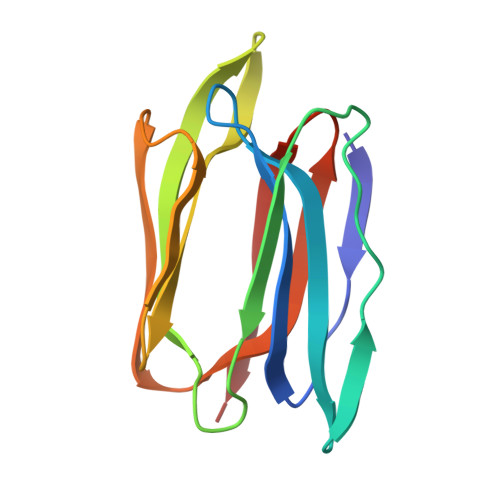
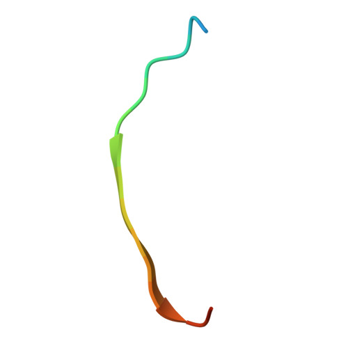
    The crystal structures of jacalin complexed with Gal α-(1,4) Gal and Gal α-(1,3) Gal β-(1,4) Gal have been determined with the primary objective of exploring the effect of linkage on the location of reducing and non-reducing sugars in the extended binding site of the lectin, an issue which has not been studied thoroughly. Contrary to the earlier surmise based on simple steric considerations, the two structures demonstrate that α-linked sugars can bind to jacalin with nonreducing sugar at the primary binding site. This is made possible substantially on account of the hitherto underestimated plasticity of a non-polar region of the extended binding site. Modeling studies involving conformational search and energy minimization, along with available crystallographic and thermodynamic data, indicate a strong preference for complexation with Gal β-(1,3) Gal with the reducing Gal at the primary site, followed by that with Gal α-(1,3) Gal, with the reducing or non-reducing Gal located at the primary binding site. This observation is in consonance with the facility of jacalin to bind mucin type O-glycans containing T-antigen core. © 2016 IUBMB Life, 68(12):971-979, 2016.


  • Organizational Affiliation

    Molecular Biophysics Unit, Indian Institute of Science, Bangalore, Karnataka, 560012, India.


Macromolecules
Find similar proteins by:  (by identity cutoff)  |  3D Structure
Entity ID: 1
MoleculeChains Sequence LengthOrganismDetailsImage
Agglutinin alpha chain
A, C, E, G
133Artocarpus integerMutation(s): 0 
UniProt
Find proteins for P18670 (Artocarpus integer)
Explore P18670 
Go to UniProtKB:  P18670
Entity Groups  
Sequence Clusters30% Identity50% Identity70% Identity90% Identity95% Identity100% Identity
UniProt GroupP18670
Sequence Annotations
Expand
  • Reference Sequence

Find similar proteins by:  Sequence   |   3D Structure  

Entity ID: 2
MoleculeChains Sequence LengthOrganismDetailsImage
Agglutinin beta-3 chain
B, D, F, H
19Artocarpus integerMutation(s): 0 
UniProt
Find proteins for P18673 (Artocarpus integer)
Explore P18673 
Go to UniProtKB:  P18673
Entity Groups  
Sequence Clusters30% Identity50% Identity70% Identity90% Identity95% Identity100% Identity
UniProt GroupP18673
Sequence Annotations
Expand
  • Reference Sequence
Oligosaccharides

Help

Entity ID: 3
MoleculeChains Length2D Diagram Glycosylation3D Interactions
alpha-D-galactopyranose-(1-4)-alpha-D-galactopyranose
I, J
2N/A
Glycosylation Resources
GlyTouCan:  G54046EC
GlyCosmos:  G54046EC
GlyGen:  G54046EC
Small Molecules
Ligands 3 Unique
IDChains Name / Formula / InChI Key2D Diagram3D Interactions
PEG
Query on PEG

Download Ideal Coordinates CCD File 
Q [auth A]DI(HYDROXYETHYL)ETHER
C4 H10 O3
MTHSVFCYNBDYFN-UHFFFAOYSA-N
EDO
Query on EDO

Download Ideal Coordinates CCD File 
AA [auth C]
BA [auth C]
DA [auth E]
EA [auth E]
FA [auth E]
AA [auth C],
BA [auth C],
DA [auth E],
EA [auth E],
FA [auth E],
GA [auth E],
HA [auth E],
IA [auth E],
JA [auth E],
K [auth A],
KA [auth E],
L [auth A],
LA [auth E],
M [auth A],
MA [auth E],
NA [auth E],
PA [auth G],
QA [auth G],
RA [auth G],
S [auth C],
SA [auth G],
T [auth C],
U [auth C],
V [auth C],
W [auth C],
X [auth C],
Y [auth C],
Z [auth C]
1,2-ETHANEDIOL
C2 H6 O2
LYCAIKOWRPUZTN-UHFFFAOYSA-N
IPA
Query on IPA

Download Ideal Coordinates CCD File 
CA [auth C]
N [auth A]
O [auth A]
OA [auth E]
P [auth A]
CA [auth C],
N [auth A],
O [auth A],
OA [auth E],
P [auth A],
R [auth B]
ISOPROPYL ALCOHOL
C3 H8 O
KFZMGEQAYNKOFK-UHFFFAOYSA-N
Experimental Data & Validation

Experimental Data

  • Method: X-RAY DIFFRACTION
  • Resolution: 1.67 Å
  • R-Value Free: 0.213 
  • R-Value Work: 0.164 
  • R-Value Observed: 0.167 
  • Space Group: P 1 21 1
Unit Cell:
Length ( Å )Angle ( ˚ )
a = 58.56α = 90
b = 81.09β = 108.05
c = 63.25γ = 90
Software Package:
Software NamePurpose
REFMACrefinement
iMOSFLMdata reduction
SCALAdata scaling
PHASERphasing

Structure Validation

View Full Validation Report


Entry History & Funding Information

Deposition Data


Funding OrganizationLocationGrant Number
Department of Science and Technology, Govt. of IndiaIndia--

Revision History  (Full details and data files)

  • Version 1.0: 2016-12-14
    Type: Initial release
  • Version 2.0: 2020-07-29
    Type: Remediation
    Reason: Carbohydrate remediation
    Changes: Atomic model, Data collection, Derived calculations, Structure summary
  • Version 2.1: 2023-11-08
    Changes: Data collection, Database references, Derived calculations, Refinement description, Structure summary