5J4T

Structure of tetrameric jacalin complexed with GlcNAc beta-(1,3) Gal-beta-OMe


Experimental Data Snapshot

  • Method: X-RAY DIFFRACTION
  • Resolution: 1.94 Å
  • R-Value Free: 0.225 
  • R-Value Work: 0.176 
  • R-Value Observed: 0.180 

wwPDB Validation   3D Report Full Report


Ligand Structure Quality Assessment 


This is version 2.1 of the entry. See complete history


Literature

Distortion of the ligand molecule as a strategy for modulating binding affinity: Further studies involving complexes of jacalin with beta-substituted disaccharides.

Abhinav, K.V.Sharma, K.Surolia, A.Vijayan, M.

(2017) IUBMB Life 69: 72-78

  • DOI: https://doi.org/10.1002/iub.1593
  • Primary Citation of Related Structures:  
    5J4T, 5J4X

  • PubMed Abstract: 

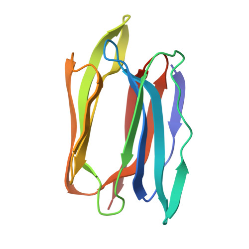
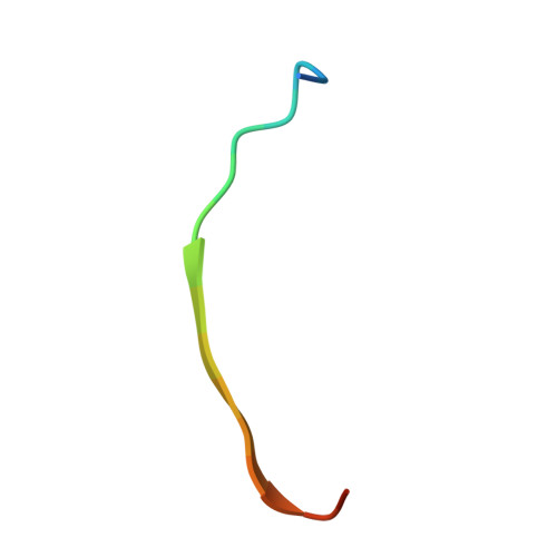
    Crystal structures of jacalin in complex with GlcNAc β-(1,3) Gal-β-OMe and Gal β-(1,3) Gal-β-OMe have been determined. The binding of the ligands to jacalin is similar to that of analogous α-substituted disaccharides. However, the β-substituted β-(1,3) linked disaccharides get distorted at the anomeric center and the glycosidic linkage. The distortion results in higher internal energies of the ligands leading to lower affinity to the lectin. This confirms the possibility of using ligand distortion as a strategy for modulating binding affinity. Unlike in the case of β-substituted monosaccharides bound to jacalin, where a larger distortion at the anomeric center was observed, smaller distortions are distributed among two centers in the structures of the two β-substituted β-(1,3) linked disaccharides presented here. These disaccharides, like the unsubstituted and α-substituted counterparts, bind jacalin with the reducing Gal at the primary binding site, indicating that the lower binding affinity of β-substituted disaccharides is not enough to overcome the intrinsic propensity of Gal β-(1,3) Gal-based disaccharides to bind jacalin with the reducing sugar at the primary site. © 2017 IUBMB Life, 69(2):72-78, 2017.


  • Organizational Affiliation

    Molecular Biophysics Unit, , Indian Institute of Science, Bangalore, India.


Macromolecules
Find similar proteins by:  (by identity cutoff)  |  3D Structure
Entity ID: 1
MoleculeChains Sequence LengthOrganismDetailsImage
Agglutinin alpha chain
A, C, E, G
133Artocarpus integerMutation(s): 0 
UniProt
Find proteins for P18670 (Artocarpus integer)
Explore P18670 
Go to UniProtKB:  P18670
Entity Groups  
Sequence Clusters30% Identity50% Identity70% Identity90% Identity95% Identity100% Identity
UniProt GroupP18670
Sequence Annotations
Expand
  • Reference Sequence

Find similar proteins by:  Sequence   |   3D Structure  

Entity ID: 2
MoleculeChains Sequence LengthOrganismDetailsImage
Agglutinin beta-3 chain
B, D, F, H
19Artocarpus integerMutation(s): 0 
UniProt
Find proteins for P18673 (Artocarpus integer)
Explore P18673 
Go to UniProtKB:  P18673
Entity Groups  
Sequence Clusters30% Identity50% Identity70% Identity90% Identity95% Identity100% Identity
UniProt GroupP18673
Sequence Annotations
Expand
  • Reference Sequence
Oligosaccharides

Help

Entity ID: 3
MoleculeChains Length2D Diagram Glycosylation3D Interactions
2-acetamido-2-deoxy-beta-D-glucopyranose-(1-3)-methyl beta-D-galactopyranoside
I
2N/A
Glycosylation Resources
GlyTouCan:  G35250SW
GlyCosmos:  G35250SW
Small Molecules
Ligands 3 Unique
IDChains Name / Formula / InChI Key2D Diagram3D Interactions
MBG
Query on MBG

Download Ideal Coordinates CCD File 
P [auth G]methyl beta-D-galactopyranoside
C7 H14 O6
HOVAGTYPODGVJG-VOQCIKJUSA-N
PEG
Query on PEG

Download Ideal Coordinates CCD File 
N [auth E]DI(HYDROXYETHYL)ETHER
C4 H10 O3
MTHSVFCYNBDYFN-UHFFFAOYSA-N
EDO
Query on EDO

Download Ideal Coordinates CCD File 
J [auth A]
K [auth A]
L [auth A]
M [auth E]
O [auth F]
J [auth A],
K [auth A],
L [auth A],
M [auth E],
O [auth F],
Q [auth G],
R [auth G],
S [auth H]
1,2-ETHANEDIOL
C2 H6 O2
LYCAIKOWRPUZTN-UHFFFAOYSA-N
Experimental Data & Validation

Experimental Data

  • Method: X-RAY DIFFRACTION
  • Resolution: 1.94 Å
  • R-Value Free: 0.225 
  • R-Value Work: 0.176 
  • R-Value Observed: 0.180 
  • Space Group: P 1 21 1
Unit Cell:
Length ( Å )Angle ( ˚ )
a = 58.31α = 90
b = 80.65β = 107.9
c = 62.85γ = 90
Software Package:
Software NamePurpose
REFMACrefinement
iMOSFLMdata reduction
SCALAdata scaling
PHASERphasing

Structure Validation

View Full Validation Report


Ligand Structure Quality Assessment 


Entry History & Funding Information

Deposition Data


Funding OrganizationLocationGrant Number
Department of Science and Technology, Govt. of IndiaIndia--

Revision History  (Full details and data files)

  • Version 1.0: 2017-02-08
    Type: Initial release
  • Version 2.0: 2020-07-29
    Type: Remediation
    Reason: Carbohydrate remediation
    Changes: Atomic model, Data collection, Derived calculations, Structure summary
  • Version 2.1: 2023-11-08
    Changes: Data collection, Database references, Derived calculations, Refinement description, Structure summary