5IN4

Crystal Structure of GDP-mannose 4,6 dehydratase bound to a GDP-fucose based inhibitor


Experimental Data Snapshot

  • Method: X-RAY DIFFRACTION
  • Resolution: 1.60 Å
  • R-Value Free: 0.176 
  • R-Value Work: 0.146 
  • R-Value Observed: 0.148 

wwPDB Validation   3D Report Full Report


Ligand Structure Quality Assessment 


This is version 1.3 of the entry. See complete history


Literature

Facile Modulation of Antibody Fucosylation with Small Molecule Fucostatin Inhibitors and Cocrystal Structure with GDP-Mannose 4,6-Dehydratase.

Allen, J.G.Mujacic, M.Frohn, M.J.Pickrell, A.J.Kodama, P.Bagal, D.San Miguel, T.Sickmier, E.A.Osgood, S.Swietlow, A.Li, V.Jordan, J.B.Kim, K.W.Rousseau, A.C.Kim, Y.J.Caille, S.Achmatowicz, M.Thiel, O.Fotsch, C.H.Reddy, P.McCarter, J.D.

(2016) ACS Chem Biol 11: 2734-2743

  • DOI: https://doi.org/10.1021/acschembio.6b00460
  • Primary Citation of Related Structures:  
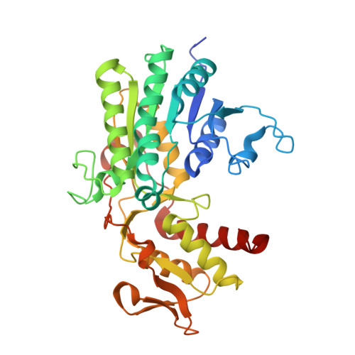
    5IN4, 5IN5

  • PubMed Abstract: 

    The efficacy of therapeutic antibodies that induce antibody-dependent cellular cytotoxicity can be improved by reduced fucosylation. Consequently, fucosylation is a critical product attribute of monoclonal antibodies produced as protein therapeutics. Small molecule fucosylation inhibitors have also shown promise as potential therapeutics in animal models of tumors, arthritis, and sickle cell disease. Potent small molecule metabolic inhibitors of cellular protein fucosylation, 6,6,6-trifluorofucose per-O-acetate and 6,6,6-trifluorofucose (fucostatin I), were identified that reduces the fucosylation of recombinantly expressed antibodies in cell culture in a concentration-dependent fashion enabling the controlled modulation of protein fucosylation levels. 6,6,6-Trifluorofucose binds at an allosteric site of GDP-mannose 4,6-dehydratase (GMD) as revealed for the first time by the X-ray cocrystal structure of a bound allosteric GMD inhibitor. 6,6,6-Trifluorofucose was found to be incorporated in place of fucose at low levels (<1%) in the glycans of recombinantly expressed antibodies. A fucose-1-phosphonate analog, fucostatin II, was designed that inhibits fucosylation with no incorporation into antibody glycans, allowing the production of afucosylated antibodies in which the incorporation of non-native sugar is completely absent-a key advantage in the production of therapeutic antibodies, especially biosimilar antibodies. Inhibitor structure-activity relationships, identification of cellular and inhibitor metabolites in inhibitor-treated cells, fucose competition studies, and the production of recombinant antibodies with varying levels of fucosylation are described.


  • Organizational Affiliation

    Therapeutic Discovery, Amgen Inc. , One Amgen Center Drive, Thousand Oaks, California 91320, United States.


Macromolecules
Find similar proteins by:  (by identity cutoff)  |  3D Structure
Entity ID: 1
MoleculeChains Sequence LengthOrganismDetailsImage
GDP-mannose 4,6 dehydratase
A, B, C, D
364Homo sapiensMutation(s): 0 
Gene Names: GMDS
EC: 4.2.1.47
UniProt & NIH Common Fund Data Resources
Find proteins for O60547 (Homo sapiens)
Explore O60547 
Go to UniProtKB:  O60547
PHAROS:  O60547
GTEx:  ENSG00000112699 
Entity Groups  
Sequence Clusters30% Identity50% Identity70% Identity90% Identity95% Identity100% Identity
UniProt GroupO60547
Sequence Annotations
Expand
  • Reference Sequence
Small Molecules
Ligands 3 Unique
IDChains Name / Formula / InChI Key2D Diagram3D Interactions
NAP
Query on NAP

Download Ideal Coordinates CCD File 
E [auth A],
I [auth B],
K [auth C],
O [auth D]
NADP NICOTINAMIDE-ADENINE-DINUCLEOTIDE PHOSPHATE
C21 H28 N7 O17 P3
XJLXINKUBYWONI-NNYOXOHSSA-N
6CK
Query on 6CK

Download Ideal Coordinates CCD File 
G [auth A],
H [auth B],
M [auth C],
N [auth D]
[(2R,3S,4R,5R)-5-(2-amino-6-oxo-1,6-dihydro-9H-purin-9-yl)-3,4-dihydroxytetrahydrofuran-2-yl]methyl (2R,3S,4R,5S,6R)-3,4,5-trihydroxy-6-(trifluoromethyl)tetrahydro-2H-pyran-2-yl dihydrogen diphosphate (non-preferred name)
C16 H22 F3 N5 O15 P2
CBLFMUIUVZLPER-KLZHJQHWSA-N
GDP
Query on GDP

Download Ideal Coordinates CCD File 
F [auth A],
J [auth B],
L [auth C],
P [auth D]
GUANOSINE-5'-DIPHOSPHATE
C10 H15 N5 O11 P2
QGWNDRXFNXRZMB-UUOKFMHZSA-N
Binding Affinity Annotations 
IDSourceBinding Affinity
6CK Binding MOAD:  5IN4 Kd: 1.10e+4 (nM) from 1 assay(s)
Experimental Data & Validation

Experimental Data

  • Method: X-RAY DIFFRACTION
  • Resolution: 1.60 Å
  • R-Value Free: 0.176 
  • R-Value Work: 0.146 
  • R-Value Observed: 0.148 
  • Space Group: P 1 21 1
Unit Cell:
Length ( Å )Angle ( ˚ )
a = 63.028α = 90
b = 140.077β = 106.24
c = 85.953γ = 90
Software Package:
Software NamePurpose
HKL-2000data scaling
PHENIXrefinement
PDB_EXTRACTdata extraction
HKL-2000data reduction
PHASERphasing

Structure Validation

View Full Validation Report


Ligand Structure Quality Assessment 


Entry History 

Deposition Data

Revision History  (Full details and data files)

  • Version 1.0: 2016-08-17
    Type: Initial release
  • Version 1.1: 2016-11-02
    Changes: Database references
  • Version 1.2: 2017-11-22
    Changes: Derived calculations, Refinement description
  • Version 1.3: 2024-03-06
    Changes: Data collection, Database references