5IDF

Cryo-EM structure of GluA2/3 AMPA receptor heterotetramer (model II)


Experimental Data Snapshot

  • Method: ELECTRON MICROSCOPY
  • Resolution: 10.3 Å
  • Aggregation State: PARTICLE 
  • Reconstruction Method: SINGLE PARTICLE 

wwPDB Validation   3D Report Full Report


This is version 1.5 of the entry. See complete history


Literature

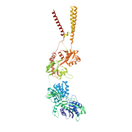
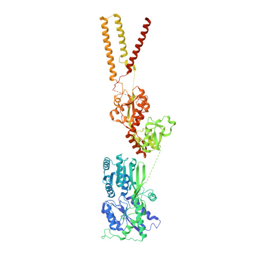
Structure and organization of heteromeric AMPA-type glutamate receptors.

Herguedas, B.Garcia-Nafria, J.Cais, O.Fernandez-Leiro, R.Krieger, J.Ho, H.Greger, I.H.

(2016) Science 352: aad3873-aad3873

  • DOI: https://doi.org/10.1126/science.aad3873
  • Primary Citation of Related Structures:  
    5FWX, 5FWY, 5IDE, 5IDF

  • PubMed Abstract: 

    AMPA-type glutamate receptors (AMPARs), which are central mediators of rapid neurotransmission and synaptic plasticity, predominantly exist as heteromers of the subunits GluA1 to GluA4. Here we report the first AMPAR heteromer structures, which deviate substantially from existing GluA2 homomer structures. Crystal structures of the GluA2/3 and GluA2/4 N-terminal domains reveal a novel compact conformation with an alternating arrangement of the four subunits around a central axis. This organization is confirmed by cysteine cross-linking in full-length receptors, and it permitted us to determine the structure of an intact GluA2/3 receptor by cryogenic electron microscopy. Two models in the ligand-free state, at resolutions of 8.25 and 10.3 angstroms, exhibit substantial vertical compression and close associations between domain layers, reminiscent of N-methyl-D-aspartate receptors. Model 1 resembles a resting state and model 2 a desensitized state, thus providing snapshots of gating transitions in the nominal absence of ligand. Our data reveal organizational features of heteromeric AMPARs and provide a framework to decipher AMPAR architecture and signaling.


  • Organizational Affiliation

    Neurobiology Division, MRC Laboratory of Molecular Biology, Cambridge, UK.


Macromolecules
Find similar proteins by:  (by identity cutoff)  |  3D Structure
Entity ID: 1
MoleculeChains Sequence LengthOrganismDetailsImage
Glutamate receptor 2
A, C
872Rattus norvegicusMutation(s): 1 
Gene Names: Gria2Glur2
Membrane Entity: Yes 
UniProt
Find proteins for P19491 (Rattus norvegicus)
Explore P19491 
Go to UniProtKB:  P19491
Entity Groups  
Sequence Clusters30% Identity50% Identity70% Identity90% Identity95% Identity100% Identity
UniProt GroupP19491
Sequence Annotations
Expand
  • Reference Sequence
Find similar proteins by:  (by identity cutoff)  |  3D Structure
Entity ID: 2
MoleculeChains Sequence LengthOrganismDetailsImage
Glutamate receptor 3
B, D
874Rattus norvegicusMutation(s): 2 
Gene Names: Gria3Glur3
Membrane Entity: Yes 
UniProt
Find proteins for P19492 (Rattus norvegicus)
Explore P19492 
Go to UniProtKB:  P19492
Entity Groups  
Sequence Clusters30% Identity50% Identity70% Identity90% Identity95% Identity100% Identity
UniProt GroupP19492
Sequence Annotations
Expand
  • Reference Sequence
Experimental Data & Validation

Experimental Data

  • Method: ELECTRON MICROSCOPY
  • Resolution: 10.3 Å
  • Aggregation State: PARTICLE 
  • Reconstruction Method: SINGLE PARTICLE 
EM Software:
TaskSoftware PackageVersion
RECONSTRUCTIONRELION

Structure Validation

View Full Validation Report


Entry History 

Deposition Data

Revision History  (Full details and data files)

  • Version 1.0: 2016-03-16
    Type: Initial release
  • Version 1.1: 2016-03-23
    Changes: Database references
  • Version 1.2: 2016-05-11
    Changes: Database references
  • Version 1.3: 2016-06-15
    Changes: Database references
  • Version 1.4: 2017-08-30
    Changes: Data collection
  • Version 1.5: 2017-10-11
    Changes: Source and taxonomy