5HMG

REFINEMENT OF THE INFLUENZA VIRUS HEMAGGLUTININ BY SIMULATED ANNEALING


Experimental Data Snapshot

  • Method: X-RAY DIFFRACTION
  • Resolution: 3.20 Å
  • R-Value Work: 0.220 

wwPDB Validation   3D Report Full Report


This is version 2.0 of the entry. See complete history


Literature

Refinement of the influenza virus hemagglutinin by simulated annealing.

Weis, W.I.Brunger, A.T.Skehel, J.J.Wiley, D.C.

(1990) J Mol Biol 212: 737-761

  • DOI: https://doi.org/10.1016/0022-2836(90)90234-D
  • Primary Citation of Related Structures:  
    2HMG, 3HMG, 4HMG, 5HMG

  • PubMed Abstract: 

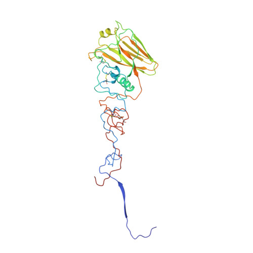
    We have applied the method of simulated annealing to the refinement of the 3 A resolution crystal structure of the influenza virus hemagglutinin glycoprotein, using the program X-PLOR. Two different methods were introduced into X-PLOR to treat the non-crystallographic symmetry present in this and in other crystal structures. In the first, only the unique protomer atoms are refined; by application of the non-crystallographic symmetry operators to the protomer atoms, the X-ray structure factor derivatives are effectively averaged, and a non-bonded energy term models the interactions of the protomer with its neighbors in the oligomer without explicit refinement of the other protomers in the crystallographic asymmetric unit. In the second method, the entire asymmetric unit is refined, but an effective energy term is added to the empirical energy that restrains symmetry-related atomic positions to their average values after least-squares superposition. Several other modifications and additions were made to previously published X-PLOR protocols, including weighting of the X-ray terms, maintenance of the temperature of the molecular dynamics simulation, treatment of charged groups, changes in the values of certain empirical energy parameters, and the use of N-linked carbohydrate empirical energy parameters. The hemagglutinin refinement proceeded in several stages. An initial round of simulated annealing of the monomer was followed by rigid-body refinement of the 3-fold non-crystallographic symmetry axis position and a second round of monomer refinement. A third round was performed on the trimer using non-crystallographic symmetry restraints in all regions except those in lattice contacts showing obvious derivations from 3-fold symmetry. The refinement was completed with several rounds of conventional positional and isotropic temperature factor refinement needed to correct bad model geometry introduced by high-temperature molecular dynamics in regions of weak electron density. This structure was then used as the basis for refinement of three crystallographically isomorphous hemagglutinin structures, including complexes with the influenza virus receptor, sialic acid. Model geometry comparable to well-refined high-resolution structures was obtained with relatively little manual intervention, demonstrating the ability of simulated annealing refinement to produce highly idealized structures at moderate resolution.


  • Organizational Affiliation

    Howard Hughes Medical Institute, Yale University, New Haven, CT 06511.


Macromolecules
Find similar proteins by:  (by identity cutoff)  |  3D Structure
Entity ID: 1
MoleculeChains Sequence LengthOrganismDetailsImage
Hemagglutinin HA1 chain
A, C, E
328Influenza A virus (A/Aichi/2/1968(H3N2))Mutation(s): 0 
Gene Names: HA
UniProt
Find proteins for P03437 (Influenza A virus (strain A/Aichi/2/1968 H3N2))
Explore P03437 
Go to UniProtKB:  P03437
Entity Groups  
Sequence Clusters30% Identity50% Identity70% Identity90% Identity95% Identity100% Identity
UniProt GroupP03437
Sequence Annotations
Expand
  • Reference Sequence
Find similar proteins by:  (by identity cutoff)  |  3D Structure
Entity ID: 2
MoleculeChains Sequence LengthOrganismDetailsImage
Hemagglutinin HA2 chain
B, D, F
175Influenza A virus (A/Aichi/2/1968(H3N2))Mutation(s): 0 
Gene Names: HA
UniProt
Find proteins for P03437 (Influenza A virus (strain A/Aichi/2/1968 H3N2))
Explore P03437 
Go to UniProtKB:  P03437
Entity Groups  
Sequence Clusters30% Identity50% Identity70% Identity90% Identity95% Identity100% Identity
UniProt GroupP03437
Sequence Annotations
Expand
  • Reference Sequence
Oligosaccharides

Help

Entity ID: 3
MoleculeChains Length2D Diagram Glycosylation3D Interactions
alpha-D-mannopyranose-(1-4)-2-acetamido-2-deoxy-beta-D-glucopyranose-(1-4)-2-acetamido-2-deoxy-beta-D-glucopyranose
G, H, I
3N-Glycosylation
Glycosylation Resources
GlyTouCan:  G62182OO
GlyCosmos:  G62182OO
GlyGen:  G62182OO
Small Molecules
Ligands 2 Unique
IDChains Name / Formula / InChI Key2D Diagram3D Interactions
SIA
Query on SIA

Download Ideal Coordinates CCD File 
J [auth A],
O [auth C],
T [auth E]
N-acetyl-alpha-neuraminic acid
C11 H19 N O9
SQVRNKJHWKZAKO-YRMXFSIDSA-N
NAG
Query on NAG

Download Ideal Coordinates CCD File 
K [auth A]
L [auth A]
M [auth A]
N [auth B]
P [auth C]
K [auth A],
L [auth A],
M [auth A],
N [auth B],
P [auth C],
Q [auth C],
R [auth C],
S [auth D],
U [auth E],
V [auth E],
W [auth E],
X [auth F]
2-acetamido-2-deoxy-beta-D-glucopyranose
C8 H15 N O6
OVRNDRQMDRJTHS-FMDGEEDCSA-N
Experimental Data & Validation

Experimental Data

  • Method: X-RAY DIFFRACTION
  • Resolution: 3.20 Å
  • R-Value Work: 0.220 
  • Space Group: P 41
Unit Cell:
Length ( Å )Angle ( ˚ )
a = 162.6α = 90
b = 162.6β = 90
c = 177.4γ = 90
Software Package:
Software NamePurpose
X-PLORrefinement

Structure Validation

View Full Validation Report


Entry History 

Deposition Data

Revision History  (Full details and data files)

  • Version 1.0: 1991-01-15
    Type: Initial release
  • Version 1.1: 2008-03-03
    Changes: Version format compliance
  • Version 1.2: 2011-07-13
    Changes: Source and taxonomy, Version format compliance
  • Version 1.3: 2017-02-01
    Changes: Structure summary
  • Version 2.0: 2020-07-29
    Type: Remediation
    Reason: Carbohydrate remediation
    Changes: Advisory, Atomic model, Data collection, Database references, Derived calculations, Other, Structure summary