5HM4

Crystal structure of oligopeptide ABC transporter, periplasmic oligopeptide-binding protein (TM1226) from THERMOTOGA MARITIMA at 2.0 A resolution


Experimental Data Snapshot

  • Method: X-RAY DIFFRACTION
  • Resolution: 2.00 Å
  • R-Value Free: 0.209 
  • R-Value Work: 0.173 
  • R-Value Observed: 0.175 

wwPDB Validation   3D Report Full Report


This is version 1.3 of the entry. See complete history


Literature

Periplasmic Binding Protein Dimer Has a Second Allosteric Event Tied to Ligand Binding.

Li, L.Ghimire-Rijal, S.Lucas, S.L.Stanley, C.B.Wright, E.Agarwal, P.K.Myles, D.A.Cuneo, M.J.

(2017) Biochemistry 56: 5328-5337

  • DOI: https://doi.org/10.1021/acs.biochem.7b00657
  • Primary Citation of Related Structures:  
    5HM4

  • PubMed Abstract: 

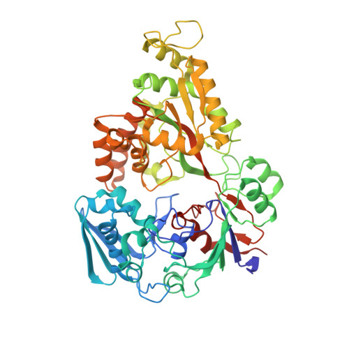
    The ligand-induced conformational changes of periplasmic binding proteins (PBP) play a key role in the acquisition of metabolites in ATP binding cassette (ABC) transport systems. This conformational change allows for differential recognition of the ligand occupancy of the PBP by the ABC transporter. This minimizes futile ATP hydrolysis in the transporter, a phenomenon in which ATP hydrolysis is not coupled to metabolite transport. In many systems, the PBP conformational change is insufficient at eliminating futile ATP hydrolysis. Here we identify an additional state of the PBP that is also allosterically regulated by the ligand. Ligand binding to the homodimeric apo PBP leads to a tightening of the interface α-helices so that the hydrogen bonding pattern shifts to that of a 3 10 helix, in-turn altering the contacts and the dynamics of the protein interface so that the monomer exists in the presence of ligand.


  • Organizational Affiliation

    Department of Biomedical Engineering, North Carolina State University , Raleigh North Carolina 27607, United States.


Macromolecules
Find similar proteins by:  (by identity cutoff)  |  3D Structure
Entity ID: 1
MoleculeChains Sequence LengthOrganismDetailsImage
Mannoside ABC transport system, sugar-binding proteinA [auth B]547Thermotoga maritimaMutation(s): 0 
Gene Names: Tmari_1233
UniProt
Find proteins for Q9X0V3 (Thermotoga maritima (strain ATCC 43589 / DSM 3109 / JCM 10099 / NBRC 100826 / MSB8))
Explore Q9X0V3 
Go to UniProtKB:  Q9X0V3
Entity Groups  
Sequence Clusters30% Identity50% Identity70% Identity90% Identity95% Identity100% Identity
UniProt GroupQ9X0V3
Sequence Annotations
Expand
  • Reference Sequence
Small Molecules
Ligands 1 Unique
IDChains Name / Formula / InChI Key2D Diagram3D Interactions
CA
Query on CA

Download Ideal Coordinates CCD File 
B
CALCIUM ION
Ca
BHPQYMZQTOCNFJ-UHFFFAOYSA-N
Experimental Data & Validation

Experimental Data

  • Method: X-RAY DIFFRACTION
  • Resolution: 2.00 Å
  • R-Value Free: 0.209 
  • R-Value Work: 0.173 
  • R-Value Observed: 0.175 
  • Space Group: P 21 2 21
Unit Cell:
Length ( Å )Angle ( ˚ )
a = 51.256α = 90
b = 65.795β = 90
c = 185.658γ = 90
Software Package:
Software NamePurpose
PHENIXrefinement
HKL-2000data reduction
HKL-2000data scaling
PHASERphasing

Structure Validation

View Full Validation Report


Entry History & Funding Information

Deposition Data


Funding OrganizationLocationGrant Number
Department of Energy (DOE, United States)United States--

Revision History  (Full details and data files)

  • Version 1.0: 2016-11-23
    Type: Initial release
  • Version 1.1: 2018-10-17
    Changes: Data collection, Database references
  • Version 1.2: 2022-03-30
    Changes: Author supporting evidence, Database references
  • Version 1.3: 2024-01-10
    Changes: Data collection, Refinement description