5HI1

Backbone Modifications in the Protein GB1 Helix: Aib24, beta-3-Lys28, beta-3-Lys31, Aib35


Experimental Data Snapshot

  • Method: X-RAY DIFFRACTION
  • Resolution: 2.15 Å
  • R-Value Free: 0.252 
  • R-Value Work: 0.214 
  • R-Value Observed: 0.217 

wwPDB Validation   3D Report Full Report


This is version 2.0 of the entry. See complete history


Literature

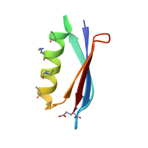
Comparison of design strategies for alpha-helix backbone modification in a protein tertiary fold.

Tavenor, N.A.Reinert, Z.E.Lengyel, G.A.Griffith, B.D.Horne, W.S.

(2016) Chem Commun (Camb) 52: 3789-3792

  • DOI: https://doi.org/10.1039/c6cc00273k
  • Primary Citation of Related Structures:  
    5HFY, 5HG2, 5HI1

  • PubMed Abstract: 

    We report here the comparison of five classes of unnatural amino acid building blocks for their ability to be accommodated into an α-helix in a protein tertiary fold context. High-resolution structural characterization and analysis of folding thermodynamics yield new insights into the relationship between backbone composition and folding energetics in α-helix mimetics and suggest refined design rules for engineering the backbones of natural sequences.


  • Organizational Affiliation

    Department of Chemistry, University of Pittsburgh, Pittsburgh, PA 15260, USA. horne@pitt.edu.


Macromolecules
Find similar proteins by:  (by identity cutoff)  |  3D Structure
Entity ID: 1
MoleculeChains Sequence LengthOrganismDetailsImage
Immunoglobulin G-binding protein G
A, B, C, D, E
A, B, C, D, E, F, G, H
57Streptococcus sp. 'group GMutation(s): 1 
UniProt
Find proteins for P19909 (Streptococcus sp. group G)
Explore P19909 
Go to UniProtKB:  P19909
Entity Groups  
Sequence Clusters30% Identity50% Identity70% Identity90% Identity95% Identity100% Identity
UniProt GroupP19909
Sequence Annotations
Expand
  • Reference Sequence
Small Molecules
Modified Residues  2 Unique
IDChains TypeFormula2D DiagramParent
AIB
Query on AIB
A, B, C, D, E
A, B, C, D, E, F, G, H
L-PEPTIDE LINKINGC4 H9 N O2ALA
B3K
Query on B3K
A, B, C, D, E
A, B, C, D, E, F, G, H
L-PEPTIDE LINKINGC7 H16 N2 O2LYS
Experimental Data & Validation

Experimental Data

  • Method: X-RAY DIFFRACTION
  • Resolution: 2.15 Å
  • R-Value Free: 0.252 
  • R-Value Work: 0.214 
  • R-Value Observed: 0.217 
  • Space Group: C 1 2 1
Unit Cell:
Length ( Å )Angle ( ˚ )
a = 74.371α = 90
b = 73.43β = 99.35
c = 79.436γ = 90
Software Package:
Software NamePurpose
PHENIXrefinement
d*TREKdata reduction
d*TREKdata scaling
PHENIXphasing

Structure Validation

View Full Validation Report


Entry History & Funding Information

Deposition Data


Funding OrganizationLocationGrant Number
National Institutes of Health/National Institute of General Medical Sciences (NIH/NIGMS)United StatesGM107161

Revision History  (Full details and data files)

  • Version 1.0: 2016-02-24
    Type: Initial release
  • Version 1.1: 2016-03-09
    Changes: Database references
  • Version 1.2: 2017-09-13
    Changes: Author supporting evidence, Derived calculations
  • Version 1.3: 2019-12-25
    Changes: Author supporting evidence
  • Version 1.4: 2023-09-27
    Changes: Data collection, Database references, Refinement description
  • Version 2.0: 2023-11-15
    Changes: Atomic model, Data collection