5H47

Crystal structure of AOL complexed with 2-MeSe-Fuc


Experimental Data Snapshot

  • Method: X-RAY DIFFRACTION
  • Resolution: 2.30 Å
  • R-Value Free: 0.258 
  • R-Value Work: 0.216 
  • R-Value Observed: 0.218 

wwPDB Validation   3D Report Full Report


Ligand Structure Quality Assessment 


This is version 1.4 of the entry. See complete history


Literature

Synthesis of seleno-fucose compounds and their application to the X-ray structural determination of carbohydrate-lectin complexes using single/multi-wavelength anomalous dispersion phasing

Shimabukuro, J.Makyio, H.Suzuki, T.Nishikawa, Y.Kawasaki, M.Imamura, A.Ishida, H.Ando, H.Kato, R.Kiso, M.

(2017) Bioorg Med Chem 25: 1132-1142

  • DOI: https://doi.org/10.1016/j.bmc.2016.12.021
  • Primary Citation of Related Structures:  
    5H47

  • PubMed Abstract: 

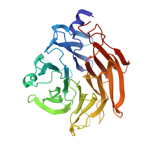
    Selenium-incorporated fucoses (seleno-fucoses) differing in the position of the seleno-substituent were synthesized and applied to the X-ray structural determination of a carbohydrate-lectin complex using single/multi-wavelength anomalous dispersion (SAD/MAD) phasing. The hydroxyl groups at the C-1, -2, -3 and -4 position of fucose were individually substituted with a methylseleno group via a transacetalization reaction using MeSeCH 2 OBn or by an S N 2 reaction with TolSe - equivalents to afford the corresponding MeSe-fucose. The three-dimensional structures of a fucose-binding lectin complexed with several of these MeSe-fucoses have been determined by SAD/MAD phasing by utilizing the diffraction of selenium in the bound MeSe-fucoses.


  • Organizational Affiliation

    Department of Applied Bioorganic Chemistry, Gifu University, 1-1 Yanagido, Gifu 501-1193, Japan; Institute for Integrated Cell-Material Sciences (WPI-iCeMS), Kyoto University, Yoshida Ushinomiya-cho, Sakyo-ku, Kyoto 606-8501, Japan.


Macromolecules
Find similar proteins by:  (by identity cutoff)  |  3D Structure
Entity ID: 1
MoleculeChains Sequence LengthOrganismDetailsImage
Uncharacterized protein
A, B, C, D, E
A, B, C, D, E, F, G, H, I, J, K, L
311Aspergillus oryzae RIB40Mutation(s): 0 
Gene Names: AO090001000189
UniProt
Find proteins for Q2UNX8 (Aspergillus oryzae (strain ATCC 42149 / RIB 40))
Explore Q2UNX8 
Go to UniProtKB:  Q2UNX8
Entity Groups  
Sequence Clusters30% Identity50% Identity70% Identity90% Identity95% Identity100% Identity
UniProt GroupQ2UNX8
Sequence Annotations
Expand
  • Reference Sequence
Small Molecules
Ligands 1 Unique
IDChains Name / Formula / InChI Key2D Diagram3D Interactions
FSW
Query on FSW

Download Ideal Coordinates CCD File 
AA [auth C]
AB [auth G]
AC [auth L]
BA [auth C]
BB [auth G]
AA [auth C],
AB [auth G],
AC [auth L],
BA [auth C],
BB [auth G],
BC [auth L],
CA [auth C],
CB [auth H],
CC [auth L],
DA [auth C],
DB [auth H],
DC [auth L],
EA [auth D],
EB [auth H],
EC [auth L],
FA [auth D],
FB [auth H],
FC [auth L],
GA [auth D],
GB [auth H],
HA [auth D],
HB [auth H],
IA [auth D],
IB [auth I],
JA [auth D],
JB [auth I],
KA [auth E],
KB [auth I],
LA [auth E],
LB [auth I],
M [auth A],
MA [auth E],
MB [auth I],
N [auth A],
NA [auth E],
NB [auth I],
O [auth A],
OA [auth E],
OB [auth J],
P [auth A],
PA [auth E],
PB [auth J],
Q [auth A],
QA [auth F],
QB [auth J],
R [auth A],
RA [auth F],
RB [auth J],
S [auth B],
SA [auth F],
SB [auth J],
T [auth B],
TA [auth F],
TB [auth J],
U [auth B],
UA [auth F],
UB [auth K],
V [auth B],
VA [auth F],
VB [auth K],
W [auth B],
WA [auth G],
WB [auth K],
X [auth B],
XA [auth G],
XB [auth K],
Y [auth C],
YA [auth G],
YB [auth K],
Z [auth C],
ZA [auth G],
ZB [auth K]
methyl 6-deoxy-2-Se-methyl-2-seleno-alpha-L-galactopyranoside
C8 H16 O4 Se
GFGVRXDXSFUDNW-FMGWEMOISA-N
Experimental Data & Validation

Experimental Data

  • Method: X-RAY DIFFRACTION
  • Resolution: 2.30 Å
  • R-Value Free: 0.258 
  • R-Value Work: 0.216 
  • R-Value Observed: 0.218 
  • Space Group: P 1 21 1
Unit Cell:
Length ( Å )Angle ( ˚ )
a = 105.972α = 90
b = 214.316β = 108.24
c = 126.531γ = 90
Software Package:
Software NamePurpose
REFMACrefinement
XDSdata reduction
Aimlessdata scaling
AutoSolphasing

Structure Validation

View Full Validation Report


Ligand Structure Quality Assessment 


Entry History 

Deposition Data

Revision History  (Full details and data files)

  • Version 1.0: 2017-01-25
    Type: Initial release
  • Version 1.1: 2017-02-15
    Changes: Database references
  • Version 1.2: 2017-08-30
    Changes: Data collection
  • Version 1.3: 2020-07-29
    Type: Remediation
    Reason: Carbohydrate remediation
    Changes: Derived calculations, Structure summary
  • Version 1.4: 2024-03-20
    Changes: Data collection, Database references, Structure summary