5GLN

Crystal structure of CoXyl43, GH43 beta-xylosidase/alpha-arabinofuranosidase from a compostmicrobial metagenome in complex with xylotriose, calcium-bound form


Experimental Data Snapshot

  • Method: X-RAY DIFFRACTION
  • Resolution: 1.70 Å
  • R-Value Free: 0.174 
  • R-Value Work: 0.158 
  • R-Value Observed: 0.159 

wwPDB Validation   3D Report Full Report


Ligand Structure Quality Assessment 


This is version 2.1 of the entry. See complete history


Literature

Crystal structure of metagenomic beta-xylosidase/ alpha-l-arabinofuranosidase activated by calcium.

Matsuzawa, T.Kaneko, S.Kishine, N.Fujimoto, Z.Yaoi, K.

(2017) J Biochem 162: 173-181

  • DOI: https://doi.org/10.1093/jb/mvx012
  • Primary Citation of Related Structures:  
    5GLK, 5GLL, 5GLM, 5GLN, 5GLO, 5GLP, 5GLQ, 5GLR

  • PubMed Abstract: 

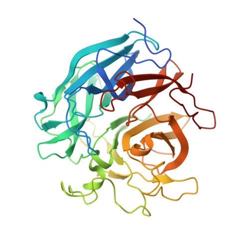
    The crystal structure of metagenomic β-xylosidase/α-l-arabinofuranosidase CoXyl43, activated by calcium ions, was determined in its apo and complexed forms with xylotriose or l-arabinose in the presence and absence of calcium. The presence of calcium ions dramatically increases the kcat of CoXyl43 for p-nitrophenyl β-d-xylopyranoside and reduces the Michaelis constant for p-nitrophenyl α-l-arabinofuranoside. CoXyl43 consists of a single catalytic domain comprised of a five-bladed β-propeller. In the presence of calcium, a single calcium ion was observed at the centre of this catalytic domain, behind the catalytic pocket. In the absence of calcium, the calcium ion was replaced with one sodium ion and one water molecule, and the positions of these cations were shifted by 1.3 Å. The histidine-319 side chain, which coordinates to the 2-hydroxyl oxygen atom of the bound xylose molecule in the catalytic pocket, also coordinates to the calcium ion, but not to the sodium ion. The calcium-dependent increase in activity appears to be caused by the structural change in the catalytic pocket induced by the tightly bound calcium ion and coordinating water molecules, and by the protonation state of glutamic acid-268, the catalytic acid of the enzyme. Our findings further elucidate the complex relationship between metal ions and glycosidases.


  • Organizational Affiliation

    Bioproduction Research Institute, National Institute of Advanced Industrial Science and Technology (AIST), Tsukuba Central 6, 1-1-1 Higashi, Tsukuba, Ibaraki 305-8566, Japan.


Macromolecules
Find similar proteins by:  (by identity cutoff)  |  3D Structure
Entity ID: 1
MoleculeChains Sequence LengthOrganismDetailsImage
Glycoside hydrolase family 43
A, B
344uncultured bacteriumMutation(s): 0 
Gene Names: coxyl43
UniProt
Find proteins for A0A0H5BL38 (uncultured bacterium)
Explore A0A0H5BL38 
Go to UniProtKB:  A0A0H5BL38
Entity Groups  
Sequence Clusters30% Identity50% Identity70% Identity90% Identity95% Identity100% Identity
UniProt GroupA0A0H5BL38
Sequence Annotations
Expand
  • Reference Sequence
Oligosaccharides

Help

Entity ID: 2
MoleculeChains Length2D Diagram Glycosylation3D Interactions
beta-D-xylopyranose-(1-4)-beta-D-xylopyranose
C, D
2N/A
Glycosylation Resources
GlyTouCan:  G87728WL
GlyCosmos:  G87728WL
Entity ID: 3
MoleculeChains Length2D Diagram Glycosylation3D Interactions
beta-D-xylopyranose-(1-4)-beta-D-xylopyranose-(1-4)-beta-D-xylopyranose
E, F
3N/A
Glycosylation Resources
GlyTouCan:  G87429QT
GlyCosmos:  G87429QT
Small Molecules
Ligands 5 Unique
IDChains Name / Formula / InChI Key2D Diagram3D Interactions
XYP
Query on XYP

Download Ideal Coordinates CCD File 
J [auth A]beta-D-xylopyranose
C5 H10 O5
SRBFZHDQGSBBOR-KKQCNMDGSA-N
XYS
Query on XYS

Download Ideal Coordinates CCD File 
I [auth A],
N [auth B]
alpha-D-xylopyranose
C5 H10 O5
SRBFZHDQGSBBOR-LECHCGJUSA-N
ACT
Query on ACT

Download Ideal Coordinates CCD File 
K [auth A],
O [auth B]
ACETATE ION
C2 H3 O2
QTBSBXVTEAMEQO-UHFFFAOYSA-M
CA
Query on CA

Download Ideal Coordinates CCD File 
G [auth A],
L [auth B]
CALCIUM ION
Ca
BHPQYMZQTOCNFJ-UHFFFAOYSA-N
NA
Query on NA

Download Ideal Coordinates CCD File 
H [auth A],
M [auth B]
SODIUM ION
Na
FKNQFGJONOIPTF-UHFFFAOYSA-N
Experimental Data & Validation

Experimental Data

  • Method: X-RAY DIFFRACTION
  • Resolution: 1.70 Å
  • R-Value Free: 0.174 
  • R-Value Work: 0.158 
  • R-Value Observed: 0.159 
  • Space Group: P 1 21 1
Unit Cell:
Length ( Å )Angle ( ˚ )
a = 74.76α = 90
b = 61.587β = 95.84
c = 78.922γ = 90
Software Package:
Software NamePurpose
REFMACrefinement
HKL-2000data reduction
SCALEPACKdata scaling
MOLREPphasing

Structure Validation

View Full Validation Report


Ligand Structure Quality Assessment 


Entry History & Funding Information

Deposition Data


Funding OrganizationLocationGrant Number
New Energy and Industrial Technology Development Organization (NEDO)Japan--

Revision History  (Full details and data files)

  • Version 1.0: 2017-03-15
    Type: Initial release
  • Version 1.1: 2017-12-06
    Changes: Data collection, Database references
  • Version 2.0: 2020-07-29
    Type: Remediation
    Reason: Carbohydrate remediation
    Changes: Atomic model, Data collection, Derived calculations, Structure summary
  • Version 2.1: 2023-11-08
    Changes: Data collection, Database references, Derived calculations, Refinement description, Structure summary