5G2C

The crystal structure of light-driven chloride pump ClR (T102D) mutant at pH 4.5.


Experimental Data Snapshot

  • Method: X-RAY DIFFRACTION
  • Resolution: 2.31 Å
  • R-Value Free: 0.239 
  • R-Value Work: 0.195 
  • R-Value Observed: 0.197 

wwPDB Validation   3D Report Full Report


Ligand Structure Quality Assessment 


This is version 1.2 of the entry. See complete history


Literature

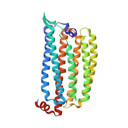
Crystal Structure and Functional Characterization of a Light-Driven Chloride Pump Having an Ntq Motif.

Kim, K.Kwon, S.Jun, S.Cha, J.S.Kim, H.Lee, W.Kim, J.F.Cho, H.

(2016) Nat Commun 7: 12677

  • DOI: https://doi.org/10.1038/ncomms12677
  • Primary Citation of Related Structures:  
    5G28, 5G2A, 5G2C, 5G2D, 5G54

  • PubMed Abstract: 

    A novel light-driven chloride-pumping rhodopsin (ClR) containing an 'NTQ motif' in its putative ion conduction pathway has been discovered and functionally characterized in a genomic analysis study of a marine bacterium. Here we report the crystal structure of ClR from the flavobacterium Nonlabens marinus S1-08(T) determined under two conditions at 2.0 and 1.56 Å resolutions. The structures reveal two chloride-binding sites, one around the protonated Schiff base and the other on a cytoplasmic loop. We identify a '3 omega motif' formed by three non-consecutive aromatic amino acids that is correlated with the B-C loop orientation. Detailed ClR structural analyses with functional studies in E. coli reveal the chloride ion transduction pathway. Our results help understand the molecular mechanism and physiological role of ClR and provide a structural basis for optogenetic applications.


  • Organizational Affiliation

    Department of Systems Biology and Division of Life Sciences, Yonsei University, 50 Yonsei-ro, Seoul 03722, Republic of Korea.


Macromolecules
Find similar proteins by:  (by identity cutoff)  |  3D Structure
Entity ID: 1
MoleculeChains Sequence LengthOrganismDetailsImage
CHLORIDE PUMPING RHODOPSIN275Nonlabens marinus S1-08Mutation(s): 1 
Membrane Entity: Yes 
UniProt
Find proteins for W8VZW3 (Nonlabens marinus S1-08)
Explore W8VZW3 
Go to UniProtKB:  W8VZW3
Entity Groups  
Sequence Clusters30% Identity50% Identity70% Identity90% Identity95% Identity100% Identity
UniProt GroupW8VZW3
Sequence Annotations
Expand
  • Reference Sequence
Small Molecules
Ligands 4 Unique
IDChains Name / Formula / InChI Key2D Diagram3D Interactions
RET
Query on RET

Download Ideal Coordinates CCD File 
I [auth A]RETINAL
C20 H28 O
NCYCYZXNIZJOKI-OVSJKPMPSA-N
OLA
Query on OLA

Download Ideal Coordinates CCD File 
B [auth A]
C [auth A]
D [auth A]
E [auth A]
F [auth A]
B [auth A],
C [auth A],
D [auth A],
E [auth A],
F [auth A],
G [auth A],
K [auth A],
L [auth A]
OLEIC ACID
C18 H34 O2
ZQPPMHVWECSIRJ-KTKRTIGZSA-N
PEG
Query on PEG

Download Ideal Coordinates CCD File 
J [auth A]DI(HYDROXYETHYL)ETHER
C4 H10 O3
MTHSVFCYNBDYFN-UHFFFAOYSA-N
CL
Query on CL

Download Ideal Coordinates CCD File 
H [auth A]CHLORIDE ION
Cl
VEXZGXHMUGYJMC-UHFFFAOYSA-M
Experimental Data & Validation

Experimental Data

  • Method: X-RAY DIFFRACTION
  • Resolution: 2.31 Å
  • R-Value Free: 0.239 
  • R-Value Work: 0.195 
  • R-Value Observed: 0.197 
  • Space Group: C 1 2 1
Unit Cell:
Length ( Å )Angle ( ˚ )
a = 103.441α = 90
b = 50.033β = 131.47
c = 77.786γ = 90
Software Package:
Software NamePurpose
REFMACrefinement
HKL-2000data reduction
SCALEPACKdata scaling
MOLREPphasing

Structure Validation

View Full Validation Report


Ligand Structure Quality Assessment 


Entry History 

Deposition Data

Revision History  (Full details and data files)

  • Version 1.0: 2016-10-19
    Type: Initial release
  • Version 1.1: 2017-03-29
    Changes: Other
  • Version 1.2: 2024-01-10
    Changes: Data collection, Database references, Derived calculations, Other, Refinement description