5FO3

ZapC cell division regulator from E. coli


Experimental Data Snapshot

  • Method: X-RAY DIFFRACTION
  • Resolution: 2.90 Å
  • R-Value Free: 0.247 
  • R-Value Work: 0.213 
  • R-Value Observed: 0.215 

wwPDB Validation   3D Report Full Report


This is version 1.2 of the entry. See complete history


Literature

Crystal Structure of the Z-Ring Associated Cell Division Protein Zapc from Escherichia Coli.

Ortiz, C.Kureisaite-Ciziene, D.Schmitz, F.Mclaughlin, S.H.Vicente, M.Lowe, J.

(2015) FEBS Lett 589: 3822

  • DOI: https://doi.org/10.1016/j.febslet.2015.11.030
  • Primary Citation of Related Structures:  
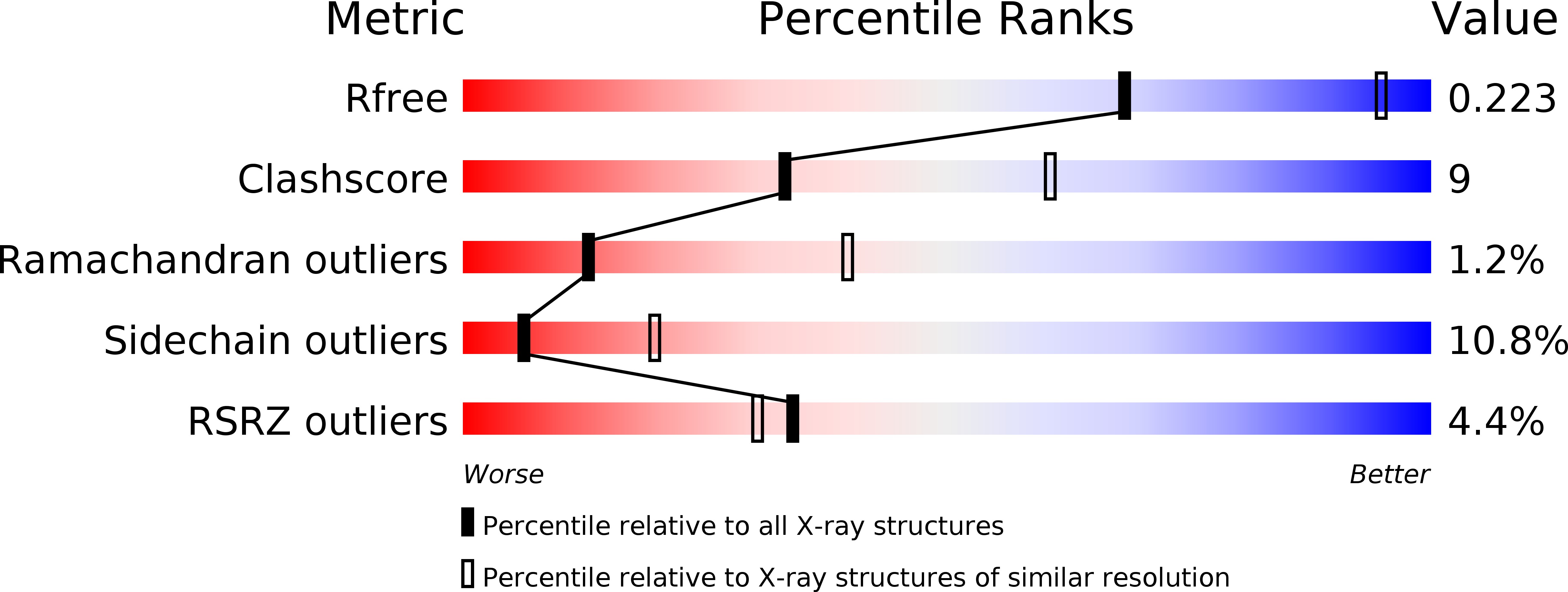
    5FO3

  • PubMed Abstract: 

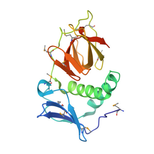
    Bacterial cell division involves a contractile ring that organises downstream proteins at the division site and which contains the tubulin homologue FtsZ. ZapC has been discovered as a non-essential regulator of FtsZ. It localises to the septal ring and deletion of zapC leads to a mild phenotype, while overexpression inhibits cell division. Interference with cell division is facilitated by an interaction with FtsZ. Here, we present the 2.9 Å crystal structure of ZapC from Escherichia coli. ZapC forms a dimer and comprises two domains that belong to the Royal superfamily of which many members bind methylated arginines or lysines. ZapC contains an N-terminal chromo-like domain and a Tudor-like C-terminal domain. We show by ITC that ZapC binds the C-terminal tail of FtsZ.


  • Organizational Affiliation

    MRC Laboratory of Molecular Biology, Francis Crick Avenue, Cambridge CB2 0QH, UK; Centro Nacional de Biotecnología, CSIC C/ Darwin 3, 28049 Madrid, Spain.


Macromolecules
Find similar proteins by:  (by identity cutoff)  |  3D Structure
Entity ID: 1
MoleculeChains Sequence LengthOrganismDetailsImage
CELL DIVISION PROTEIN ZAPC
A, B
180Escherichia coliMutation(s): 0 
UniProt
Find proteins for P75862 (Escherichia coli (strain K12))
Explore P75862 
Go to UniProtKB:  P75862
Entity Groups  
Sequence Clusters30% Identity50% Identity70% Identity90% Identity95% Identity100% Identity
UniProt GroupP75862
Sequence Annotations
Expand
  • Reference Sequence
Small Molecules
Modified Residues  1 Unique
IDChains TypeFormula2D DiagramParent
MSE
Query on MSE
A, B
L-PEPTIDE LINKINGC5 H11 N O2 SeMET
Experimental Data & Validation

Experimental Data

  • Method: X-RAY DIFFRACTION
  • Resolution: 2.90 Å
  • R-Value Free: 0.247 
  • R-Value Work: 0.213 
  • R-Value Observed: 0.215 
  • Space Group: P 43 21 2
Unit Cell:
Length ( Å )Angle ( ˚ )
a = 87.371α = 90
b = 87.371β = 90
c = 118.311γ = 90
Software Package:
Software NamePurpose
REFMACrefinement
XDSdata reduction
SCALAdata scaling
PHASERphasing

Structure Validation

View Full Validation Report


Entry History 

Deposition Data

Revision History  (Full details and data files)

  • Version 1.0: 2015-11-25
    Type: Initial release
  • Version 1.1: 2015-12-16
    Changes: Database references
  • Version 1.2: 2016-01-13
    Changes: Database references