5FF1

Two way mode of binding of antithyroid drug methimazole to mammalian heme peroxidases: Structure of the complex of lactoperoxidase with methimazole at 1.97 Angstrom resolution


Experimental Data Snapshot

  • Method: X-RAY DIFFRACTION
  • Resolution: 1.97 Å
  • R-Value Free: 0.226 
  • R-Value Work: 0.177 
  • R-Value Observed: 0.178 

wwPDB Validation   3D Report Full Report


Ligand Structure Quality Assessment 


This is version 2.1 of the entry. See complete history


Literature

Dual binding mode of antithyroid drug methimazole to mammalian heme peroxidases - structural determination of the lactoperoxidase-methimazole complex at 1.97 angstrom resolution.

Singh, R.P.Singh, A.Sirohi, H.V.Singh, A.K.Kaur, P.Sharma, S.Singh, T.P.

(2016) FEBS Open Bio 6: 640-650

  • DOI: https://doi.org/10.1002/2211-5463.12051
  • Primary Citation of Related Structures:  
    5FF1

  • PubMed Abstract: 

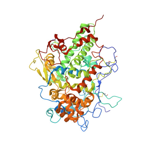
    Lactoperoxidase (LPO, EC 1.11.1.7) is a member of the mammalian heme peroxidase family which also includes thyroid peroxidase (TPO). These two enzymes have a sequence homology of 76%. The structure of LPO is known but not that of TPO. In order to determine the mode of binding of antithyroid drugs to thyroid peroxidase, we have determined the crystal structure of LPO complexed with an antithyroid drug, methimazole (MMZ) at 1.97 Å resolution. LPO was isolated from caprine colostrum, purified to homogeneity and crystallized with 20% poly(ethylene glycol)-3350. Crystals of LPO were soaked in a reservoir solution containing MMZ. The structure determination showed the presence of two crystallographically independent molecules in the asymmetric unit. Both molecules contained one molecule of MMZ, but with different orientations. MMZ was held tightly between the heme moiety on one side and the hydrophobic parts of the side chains of Arg255, Glu258, and Leu262 on the opposite side. The back of the cleft contained the side chains of Gln105 and His109 which also interacted with MMZ. In both orientations, MMZ had identical buried areas and formed a similar number of interactions. It appears that the molecules of MMZ can enter the substrate-binding channel of LPO in two opposite orientations. But once they reach the distal heme pocket, their orientations are frozen due to equally tight packing of MMZ in both orientations. This is a novel example of an inhibitor binding to an enzyme with two orientations at the same site with nearly equal occupancies.


  • Organizational Affiliation

    Department of Biophysics All India Institute of Medical Sciences New Delhi India.


Macromolecules
Find similar proteins by:  (by identity cutoff)  |  3D Structure
Entity ID: 1
MoleculeChains Sequence LengthOrganismDetailsImage
Lactoperoxidase
A, B
595Capra hircusMutation(s): 0 
UniProt
Find proteins for A0A452E9Y6 (Capra hircus)
Explore A0A452E9Y6 
Go to UniProtKB:  A0A452E9Y6
Entity Groups  
Sequence Clusters30% Identity50% Identity70% Identity90% Identity95% Identity100% Identity
UniProt GroupA0A452E9Y6
Sequence Annotations
Expand
  • Reference Sequence
Oligosaccharides

Help

Entity ID: 2
MoleculeChains Length2D Diagram Glycosylation3D Interactions
2-acetamido-2-deoxy-beta-D-glucopyranose-(1-4)-2-acetamido-2-deoxy-beta-D-glucopyranose
C
2N-Glycosylation
Glycosylation Resources
GlyTouCan:  G42666HT
GlyCosmos:  G42666HT
GlyGen:  G42666HT
Small Molecules
Ligands 6 Unique
IDChains Name / Formula / InChI Key2D Diagram3D Interactions
HEM
Query on HEM

Download Ideal Coordinates CCD File 
J [auth A],
Y [auth B]
PROTOPORPHYRIN IX CONTAINING FE
C34 H32 Fe N4 O4
KABFMIBPWCXCRK-RGGAHWMASA-L
NAG
Query on NAG

Download Ideal Coordinates CCD File 
F [auth A]
G [auth A]
H [auth A]
I [auth A]
V [auth B]
F [auth A],
G [auth A],
H [auth A],
I [auth A],
V [auth B],
W [auth B],
X [auth B]
2-acetamido-2-deoxy-beta-D-glucopyranose
C8 H15 N O6
OVRNDRQMDRJTHS-FMDGEEDCSA-N
MMZ
Query on MMZ

Download Ideal Coordinates CCD File 
D [auth A],
E [auth A],
EA [auth B],
U [auth B]
1-METHYL-1,3-DIHYDRO-2H-IMIDAZOLE-2-THIONE
C4 H6 N2 S
PMRYVIKBURPHAH-UHFFFAOYSA-N
GOL
Query on GOL

Download Ideal Coordinates CCD File 
FA [auth B]
GA [auth B]
HA [auth B]
IA [auth B]
Q [auth A]
FA [auth B],
GA [auth B],
HA [auth B],
IA [auth B],
Q [auth A],
R [auth A],
S [auth A],
T [auth A]
GLYCEROL
C3 H8 O3
PEDCQBHIVMGVHV-UHFFFAOYSA-N
NO3
Query on NO3

Download Ideal Coordinates CCD File 
AA [auth B]
BA [auth B]
CA [auth B]
DA [auth B]
L [auth A]
AA [auth B],
BA [auth B],
CA [auth B],
DA [auth B],
L [auth A],
M [auth A],
N [auth A],
O [auth A],
P [auth A]
NITRATE ION
N O3
NHNBFGGVMKEFGY-UHFFFAOYSA-N
CA
Query on CA

Download Ideal Coordinates CCD File 
K [auth A],
Z [auth B]
CALCIUM ION
Ca
BHPQYMZQTOCNFJ-UHFFFAOYSA-N
Binding Affinity Annotations 
IDSourceBinding Affinity
MMZ BindingDB:  5FF1 IC50: 8400 (nM) from 1 assay(s)
Experimental Data & Validation

Experimental Data

  • Method: X-RAY DIFFRACTION
  • Resolution: 1.97 Å
  • R-Value Free: 0.226 
  • R-Value Work: 0.177 
  • R-Value Observed: 0.178 
  • Space Group: P 1 21 1
Unit Cell:
Length ( Å )Angle ( ˚ )
a = 80.31α = 90
b = 93.02β = 89.97
c = 81.53γ = 90
Software Package:
Software NamePurpose
REFMACrefinement
HKL-2000data reduction
SCALEPACKdata scaling
AMoREphasing

Structure Validation

View Full Validation Report


Ligand Structure Quality Assessment 


Entry History 

Deposition Data

Revision History  (Full details and data files)

  • Version 1.0: 2016-01-13
    Type: Initial release
  • Version 1.1: 2016-07-27
    Changes: Database references
  • Version 2.0: 2020-07-29
    Type: Remediation
    Reason: Carbohydrate remediation
    Changes: Atomic model, Data collection, Derived calculations, Structure summary
  • Version 2.1: 2023-11-08
    Changes: Data collection, Database references, Refinement description, Structure summary