5FAE

N184K pathological variant of gelsolin domain 2 (trigonal form)


Experimental Data Snapshot

  • Method: X-RAY DIFFRACTION
  • Resolution: 1.70 Å
  • R-Value Free: 0.225 
  • R-Value Work: 0.182 
  • R-Value Observed: 0.186 

wwPDB Validation   3D Report Full Report


This is version 1.1 of the entry. See complete history


Literature

Molecular basis of a novel renal amyloidosis due to N184K gelsolin variant.

Boni, F.Milani, M.Porcari, R.Barbiroli, A.Ricagno, S.de Rosa, M.

(2016) Sci Rep 6: 33463-33463

  • DOI: https://doi.org/10.1038/srep33463
  • Primary Citation of Related Structures:  
    5FAE, 5FAF

  • PubMed Abstract: 

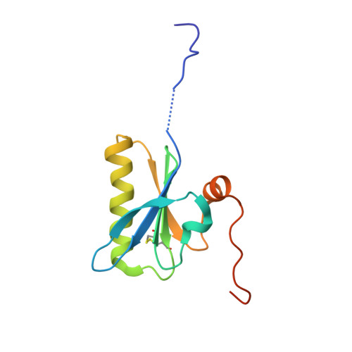
    Mutations in gelsolin are responsible for a systemic amyloidosis first described in 1969. Until recently, the disease was associated with two substitutions of the same residue, leading to the loss of the calcium binding site. Novel interest arose in 2014 when the N184K variant of the protein was identified as the etiological agent of a novel kidney-localized amyloidosis. Here we provide a first rationale for N184K pathogenicity. We show that the mutation induces a destabilization of gelsolin second domain, without compromising its calcium binding capacity. X-ray data combined with molecular dynamics simulations demonstrates that the primary source of the destabilization is a loss of connectivity in proximity of the metal. Such rearrangement of the H-bond network does not have a major impact on the overall fold of the domain, nevertheless, it increases the flexibility of a stretch of the protein, which is consequently processed by furin protease. Overall our data suggest that the N184K variant is subjected to the same aberrant proteolytic events responsible for the formation of amyloidogenic fragments in the previously characterized mutants. At the same time our data suggest that a broader number of mutations, unrelated to the metal binding site, can lead to a pathogenic phenotype.


  • Organizational Affiliation

    CNR Istituto di Biofisica, c/o Dipartimento di Bioscienze, Università degli Studi di Milano, 20133 Milano, Italy.


Macromolecules
Find similar proteins by:  (by identity cutoff)  |  3D Structure
Entity ID: 1
MoleculeChains Sequence LengthOrganismDetailsImage
Gelsolin119Homo sapiensMutation(s): 1 
Gene Names: GSN
UniProt & NIH Common Fund Data Resources
Find proteins for P06396 (Homo sapiens)
Explore P06396 
Go to UniProtKB:  P06396
PHAROS:  P06396
GTEx:  ENSG00000148180 
Entity Groups  
Sequence Clusters30% Identity50% Identity70% Identity90% Identity95% Identity100% Identity
UniProt GroupP06396
Sequence Annotations
Expand
  • Reference Sequence
Small Molecules
Ligands 4 Unique
IDChains Name / Formula / InChI Key2D Diagram3D Interactions
PEG
Query on PEG

Download Ideal Coordinates CCD File 
G [auth A]DI(HYDROXYETHYL)ETHER
C4 H10 O3
MTHSVFCYNBDYFN-UHFFFAOYSA-N
SO4
Query on SO4

Download Ideal Coordinates CCD File 
C [auth A],
D [auth A],
E [auth A],
F [auth A]
SULFATE ION
O4 S
QAOWNCQODCNURD-UHFFFAOYSA-L
CA
Query on CA

Download Ideal Coordinates CCD File 
B [auth A]CALCIUM ION
Ca
BHPQYMZQTOCNFJ-UHFFFAOYSA-N
CL
Query on CL

Download Ideal Coordinates CCD File 
H [auth A]CHLORIDE ION
Cl
VEXZGXHMUGYJMC-UHFFFAOYSA-M
Experimental Data & Validation

Experimental Data

  • Method: X-RAY DIFFRACTION
  • Resolution: 1.70 Å
  • R-Value Free: 0.225 
  • R-Value Work: 0.182 
  • R-Value Observed: 0.186 
  • Space Group: P 31 2 1
Unit Cell:
Length ( Å )Angle ( ˚ )
a = 46.919α = 90
b = 46.919β = 90
c = 84.623γ = 120
Software Package:
Software NamePurpose
PHENIXrefinement
PHASERphasing
MOSFLMdata reduction
Aimlessdata scaling

Structure Validation

View Full Validation Report


Entry History & Funding Information

Deposition Data


Funding OrganizationLocationGrant Number
TelethonItalyGEP15070

Revision History  (Full details and data files)

  • Version 1.0: 2016-10-05
    Type: Initial release
  • Version 1.1: 2024-01-10
    Changes: Data collection, Database references, Refinement description