Acetylation of Surface Lysine Groups of a Protein Alters the Organization and Composition of Its Crystal Contacts.
Kang, K., Choi, J.M., Fox, J.M., Snyder, P.W., Moustakas, D.T., Whitesides, G.M.(2016) J Phys Chem B 120: 6461-6468
- PubMed: 27292012
- DOI: https://doi.org/10.1021/acs.jpcb.6b01105
- Primary Citation of Related Structures:
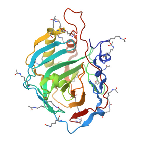
5EZT - PubMed Abstract:
This paper uses crystals of bovine carbonic anhydrase (CA) and its acetylated variant to examine (i) how a large negative formal charge can be accommodated in protein-protein interfaces, (ii) why lysine residues are often excluded from them, and (iii) how changes in the surface charge of a protein can alter the structure and organization of protein-protein interfaces. It demonstrates that acetylation of lysine residues on the surface of CA increases the participation of polar residues (particularly acetylated lysine) in protein-protein interfaces, and decreases the participation of nonpolar residues in those interfaces. Negatively charged residues are accommodated in protein-protein interfaces via (i) hydrogen bonds or van der Waals interactions with polar residues or (ii) salt bridges with other charged residues. The participation of acetylated lysine in protein-protein interfaces suggests that unacetylated lysine tends to be excluded from interfaces because of its positive charge, and not because of a loss in conformational entropy. Results also indicate that crystal contacts in acetylated CA become less constrained geometrically and, as a result, more closely packed (i.e., more tightly clustered spatially) than those of native CA. This study demonstrates a physical-organic approach-and a well-defined model system-for studying the role of charges in protein-protein interactions.
Organizational Affiliation:
Department of Chemistry and Chemical Biology, Harvard University , 12 Oxford Street, Cambridge, Massachusetts 02138, United States.