5EO8

Crystal structure of AOL(868)


Experimental Data Snapshot

  • Method: X-RAY DIFFRACTION
  • Resolution: 1.60 Å
  • R-Value Free: 0.183 
  • R-Value Work: 0.164 
  • R-Value Observed: 0.165 

wwPDB Validation   3D Report Full Report


Ligand Structure Quality Assessment 


This is version 2.1 of the entry. See complete history


Literature

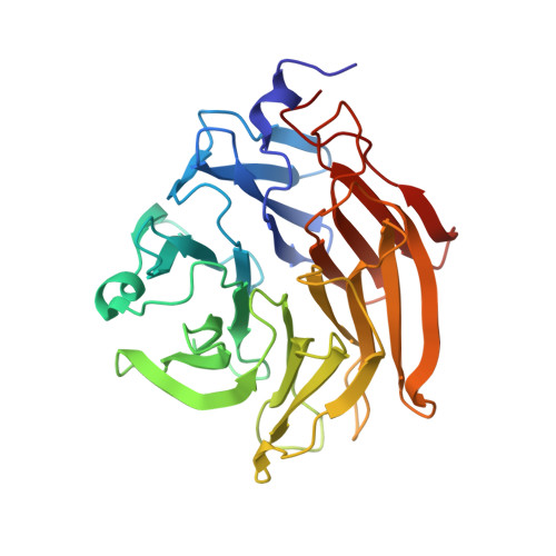
Six independent fucose-binding sites in the crystal structure of Aspergillus oryzae lectin

Makyio, H.Shimabukuro, J.Suzuki, T.Imamura, A.Ishida, H.Kiso, M.Ando, H.Kato, R.

(2016) Biochem Biophys Res Commun 477: 477-482

  • DOI: https://doi.org/10.1016/j.bbrc.2016.06.069
  • Primary Citation of Related Structures:  
    5EO7, 5EO8

  • PubMed Abstract: 

    The crystal structure of AOL (a fucose-specific lectin of Aspergillus oryzae) has been solved by SAD (single-wavelength anomalous diffraction) and MAD (multi-wavelength anomalous diffraction) phasing of seleno-fucosides. The overall structure is a six-bladed β-propeller similar to that of other fucose-specific lectins. The fucose moieties of the seleno-fucosides are located in six fucose-binding sites. Although the Arg and Glu/Gln residues bound to the fucose moiety are common to all fucose-binding sites, the amino-acid residues involved in fucose binding at each site are not identical. The varying peak heights of the seleniums in the electron density map suggest that each fucose-binding site has a different carbohydrate binding affinity.


  • Organizational Affiliation

    Structural Biology Research Center, Photon Factory, Institute of Materials Structure Science, High Energy Accelerator Research Organization (KEK), 1-1 Oho, Tsukuba, Ibaraki, 305-0801, Japan.


Macromolecules
Find similar proteins by:  (by identity cutoff)  |  3D Structure
Entity ID: 1
MoleculeChains Sequence LengthOrganismDetailsImage
Predicted protein311Aspergillus oryzae RIB40Mutation(s): 0 
Gene Names: AO090001000189
UniProt
Find proteins for Q2UNX8 (Aspergillus oryzae (strain ATCC 42149 / RIB 40))
Explore Q2UNX8 
Go to UniProtKB:  Q2UNX8
Entity Groups  
Sequence Clusters30% Identity50% Identity70% Identity90% Identity95% Identity100% Identity
UniProt GroupQ2UNX8
Sequence Annotations
Expand
  • Reference Sequence
Experimental Data & Validation

Experimental Data

  • Method: X-RAY DIFFRACTION
  • Resolution: 1.60 Å
  • R-Value Free: 0.183 
  • R-Value Work: 0.164 
  • R-Value Observed: 0.165 
  • Space Group: P 61 2 2
Unit Cell:
Length ( Å )Angle ( ˚ )
a = 58.595α = 90
b = 58.595β = 90
c = 397.601γ = 120
Software Package:
Software NamePurpose
Aimlessdata scaling
SHELXphasing
PHENIXrefinement
PDB_EXTRACTdata extraction
XDSdata reduction

Structure Validation

View Full Validation Report


Ligand Structure Quality Assessment 


Entry History 

Deposition Data

Revision History  (Full details and data files)

  • Version 1.0: 2016-06-29
    Type: Initial release
  • Version 1.1: 2016-08-03
    Changes: Database references
  • Version 1.2: 2020-02-19
    Changes: Data collection, Database references, Derived calculations
  • Version 2.0: 2020-07-29
    Type: Remediation
    Reason: Carbohydrate remediation
    Changes: Atomic model, Derived calculations, Structure summary
  • Version 2.1: 2024-03-20
    Changes: Data collection, Database references, Structure summary