5EIK

Structure of a Trimeric Intracellular Cation channel from C. elegans in the absence of Ca2+


Experimental Data Snapshot

  • Method: X-RAY DIFFRACTION
  • Resolution: 2.30 Å
  • R-Value Free: 0.249 
  • R-Value Work: 0.214 

wwPDB Validation   3D Report Full Report


Ligand Structure Quality Assessment 


This is version 2.0 of the entry. See complete history


Literature

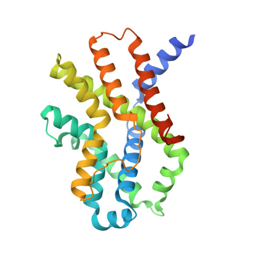
Pore architecture of TRIC channels and insights into their gating mechanism.

Yang, H.T.Hu, M.H.Guo, J.L.Ou, X.M.Cai, T.X.Liu, Z.F.

(2016) Nature 538: 537-541

  • DOI: https://doi.org/10.1038/nature19767
  • Primary Citation of Related Structures:  
    5EGI, 5EIK

  • PubMed Abstract: 

    Intracellular Ca 2+ signalling processes are fundamental to muscle contraction, neurotransmitter release, cell growth and apoptosis. Release of Ca 2+ from the intracellular stores is supported by a series of ion channels in sarcoplasmic or endoplasmic reticulum (SR/ER). Among them, two isoforms of the trimeric intracellular cation (TRIC) channel family, named TRIC-A and TRIC-B, modulate the release of Ca 2+ through the ryanodine receptor or inositol triphosphate receptor, and maintain the homeostasis of ions within SR/ER lumen. Genetic ablations or mutations of TRIC channels are associated with hypertension, heart disease, respiratory defects and brittle bone disease. Despite the pivotal function of TRIC channels in Ca 2+ signalling, their pore architectures and gating mechanisms remain unknown. Here we present the structures of TRIC-B1 and TRIC-B2 channels from Caenorhabditis elegans in complex with endogenous phosphatidylinositol-4,5-biphosphate (PtdIns(4,5)P 2 , also known as PIP 2 ) lipid molecules. The TRIC-B1/B2 proteins and PIP 2 assemble into a symmetrical homotrimeric complex. Each monomer contains an hourglass-shaped hydrophilic pore contained within a seven-transmembrane-helix domain. Structural and functional analyses unravel the central role of PIP 2 in stabilizing the cytoplasmic gate of the ion permeation pathway and reveal a marked Ca 2+ -induced conformational change in a cytoplasmic loop above the gate. A mechanistic model has been proposed to account for the complex gating mechanism of TRIC channels.


  • Organizational Affiliation

    National Laboratory of Biomacromolecules, CAS Center for Excellence in Biomacromolecules, Institute of Biophysics, Chinese Academy of Sciences, Beijing 100101, China.


Macromolecules
Find similar proteins by:  (by identity cutoff)  |  3D Structure
Entity ID: 1
MoleculeChains Sequence LengthOrganismDetailsImage
Uncharacterized protein Y57A10A.28258Caenorhabditis elegansMutation(s): 0 
Gene Names: CELE_Y57A10A.28Y57A10A.28
Membrane Entity: Yes 
UniProt
Find proteins for Q9NA73 (Caenorhabditis elegans)
Explore Q9NA73 
Go to UniProtKB:  Q9NA73
Entity Groups  
Sequence Clusters30% Identity50% Identity70% Identity90% Identity95% Identity100% Identity
UniProt GroupQ9NA73
Sequence Annotations
Expand
  • Reference Sequence
Small Molecules
Ligands 5 Unique
IDChains Name / Formula / InChI Key2D Diagram3D Interactions
PT5
Query on PT5

Download Ideal Coordinates CCD File 
B [auth A][(2R)-1-octadecanoyloxy-3-[oxidanyl-[(1R,2R,3S,4R,5R,6S)-2,3,6-tris(oxidanyl)-4,5-diphosphonooxy-cyclohexyl]oxy-phospho ryl]oxy-propan-2-yl] (8Z)-icosa-5,8,11,14-tetraenoate
C47 H85 O19 P3
CNWINRVXAYPOMW-HJBQCNPJSA-N
CPL
Query on CPL

Download Ideal Coordinates CCD File 
C [auth A]1-PALMITOYL-2-LINOLEOYL-SN-GLYCERO-3-PHOSPHOCHOLINE
C42 H80 N O8 P
JLPULHDHAOZNQI-ZTIMHPMXSA-N
DMU
Query on DMU

Download Ideal Coordinates CCD File 
J [auth A],
K [auth A],
L [auth A],
M [auth A]
DECYL-BETA-D-MALTOPYRANOSIDE
C22 H42 O11
WOQQAWHSKSSAGF-WXFJLFHKSA-N
ACT
Query on ACT

Download Ideal Coordinates CCD File 
E [auth A],
F [auth A],
G [auth A],
H [auth A],
I [auth A]
ACETATE ION
C2 H3 O2
QTBSBXVTEAMEQO-UHFFFAOYSA-M
MG
Query on MG

Download Ideal Coordinates CCD File 
D [auth A]MAGNESIUM ION
Mg
JLVVSXFLKOJNIY-UHFFFAOYSA-N
Experimental Data & Validation

Experimental Data

  • Method: X-RAY DIFFRACTION
  • Resolution: 2.30 Å
  • R-Value Free: 0.249 
  • R-Value Work: 0.214 
  • Space Group: H 3 2
Unit Cell:
Length ( Å )Angle ( ˚ )
a = 126.34α = 90
b = 126.34β = 90
c = 135.57γ = 120
Software Package:
Software NamePurpose
CNSrefinement
MOSFLMdata reduction
SCALAdata scaling
PHASERphasing

Structure Validation

View Full Validation Report


Ligand Structure Quality Assessment 


Entry History & Funding Information

Deposition Data


Funding OrganizationLocationGrant Number
National 973 projectChina2014CB910301
Chinese Academy of SciencesChinaXDB08020302

Revision History  (Full details and data files)

  • Version 1.0: 2016-10-05
    Type: Initial release
  • Version 1.1: 2019-12-25
    Changes: Database references, Derived calculations
  • Version 2.0: 2023-11-08
    Changes: Atomic model, Data collection, Database references, Derived calculations, Refinement description, Structure summary