5E7P

Crystal Structure of MSMEG_0858 (Uniprot A0QQS4), a AAA ATPase.


Experimental Data Snapshot

  • Method: X-RAY DIFFRACTION
  • Resolution: 2.51 Å
  • R-Value Free: 0.251 
  • R-Value Work: 0.206 
  • R-Value Observed: 0.208 

wwPDB Validation   3D Report Full Report


This is version 1.2 of the entry. See complete history


Literature

Crystal Structure and Biochemical Characterization of a Mycobacterium smegmatis AAA-Type Nucleoside Triphosphatase Phosphohydrolase (Msm0858).

Unciuleac, M.C.Smith, P.C.Shuman, S.

(2016) J Bacteriol 198: 1521-1533

  • DOI: https://doi.org/10.1128/JB.00905-15
  • Primary Citation of Related Structures:  
    5E7P

  • PubMed Abstract: 

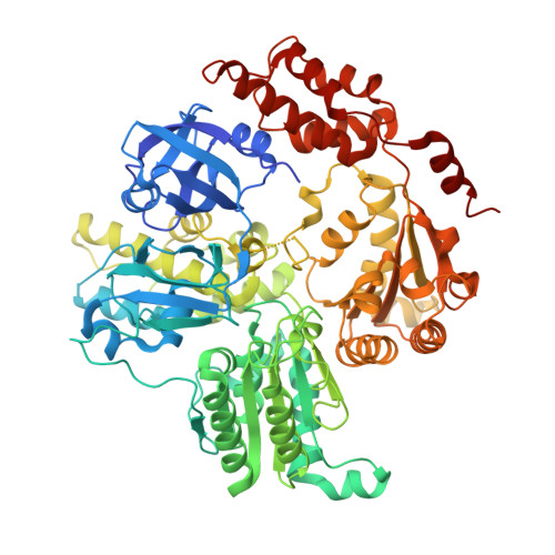
    AAA proteins (ATPases associated with various cellular activities) use the energy of ATP hydrolysis to drive conformational changes in diverse macromolecular targets. Here, we report the biochemical characterization and 2.5-Å crystal structure of a Mycobacterium smegmatis AAA protein Msm0858, the ortholog of Mycobacterium tuberculosis Rv0435c. Msm0858 is a magnesium-dependent ATPase and is active with all nucleoside triphosphates (NTPs) and deoxynucleoside triphosphates (dNTPs) as substrates. The Msm0858 structure comprises (i) an N-terminal domain (amino acids [aa] 17 to 201) composed of two β-barrel modules and (ii) two AAA domains, D1 (aa 212 to 473) and D2 (aa 476 to 744), each of which has ADP in the active site. Msm0858-ADP is a monomer in solution and in crystallized form. Msm0858 domains are structurally homologous to the corresponding modules of mammalian p97. However, the position of the N-domain modules relative to the AAA domains in the Msm0858-ADP tertiary structure is different and would impede the formation of a p97-like hexameric quaternary structure. Mutational analysis of the A-box and B-box motifs indicated that the D1 and D2 AAA domains are both capable of ATP hydrolysis. Simultaneous mutations of the D1 and D2 active-site motifs were required to abolish ATPase activity. ATPase activity was effaced by mutation of the putative D2 arginine finger, suggesting that Msm0858 might oligomerize during the ATPase reaction cycle. A truncated variant Msm0858 (aa 212 to 745) that lacks the N domain was characterized as a catalytically active homodimer. Recent studies have underscored the importance of AAA proteins (ATPases associated with various cellular activities) in the physiology of mycobacteria. This study reports the ATPase activity and crystal structure of a previously uncharacterized mycobacterial AAA protein, Msm0858. Msm0858 consists of an N-terminal β-barrel domain and two AAA domains, each with ADP bound in the active site. Msm0858 is a structural homolog of mammalian p97, with respect to the linear order and tertiary structures of their domains.


  • Organizational Affiliation

    Molecular Biology Program, Sloan-Kettering Institute, New York, New York, USA.


Macromolecules
Find similar proteins by:  (by identity cutoff)  |  3D Structure
Entity ID: 1
MoleculeChains Sequence LengthOrganismDetailsImage
Cell division control protein Cdc48
A, B
745Mycolicibacterium smegmatis MC2 155Mutation(s): 0 
Gene Names: MSMEG_0858MSMEI_0839
EC: 3.6.4.6
UniProt
Find proteins for A0QQS4 (Mycolicibacterium smegmatis (strain ATCC 700084 / mc(2)155))
Explore A0QQS4 
Go to UniProtKB:  A0QQS4
Entity Groups  
Sequence Clusters30% Identity50% Identity70% Identity90% Identity95% Identity100% Identity
UniProt GroupA0QQS4
Sequence Annotations
Expand
  • Reference Sequence
Small Molecules
Ligands 4 Unique
IDChains Name / Formula / InChI Key2D Diagram3D Interactions
ADP
Query on ADP

Download Ideal Coordinates CCD File 
C [auth A],
D [auth A],
H [auth B],
I [auth B]
ADENOSINE-5'-DIPHOSPHATE
C10 H15 N5 O10 P2
XTWYTFMLZFPYCI-KQYNXXCUSA-N
PG4
Query on PG4

Download Ideal Coordinates CCD File 
E [auth A]TETRAETHYLENE GLYCOL
C8 H18 O5
UWHCKJMYHZGTIT-UHFFFAOYSA-N
PGE
Query on PGE

Download Ideal Coordinates CCD File 
J [auth B],
K [auth B]
TRIETHYLENE GLYCOL
C6 H14 O4
ZIBGPFATKBEMQZ-UHFFFAOYSA-N
GOL
Query on GOL

Download Ideal Coordinates CCD File 
F [auth A],
G [auth A],
L [auth B]
GLYCEROL
C3 H8 O3
PEDCQBHIVMGVHV-UHFFFAOYSA-N
Experimental Data & Validation

Experimental Data

  • Method: X-RAY DIFFRACTION
  • Resolution: 2.51 Å
  • R-Value Free: 0.251 
  • R-Value Work: 0.206 
  • R-Value Observed: 0.208 
  • Space Group: P 21 21 21
Unit Cell:
Length ( Å )Angle ( ˚ )
a = 86.801α = 90
b = 111.816β = 90
c = 151.705γ = 90
Software Package:
Software NamePurpose
PHENIXrefinement
HKL-2000data reduction
HKL-2000data scaling
PHASERphasing

Structure Validation

View Full Validation Report


Entry History & Funding Information

Deposition Data


Funding OrganizationLocationGrant Number
National Institutes of Health/National Institute Of Allergy and Infectious Diseases (NIH/NIAID)United StatesAI64693

Revision History  (Full details and data files)

  • Version 1.0: 2016-08-24
    Type: Initial release
  • Version 1.1: 2017-09-13
    Changes: Author supporting evidence, Database references, Derived calculations
  • Version 1.2: 2022-04-13
    Changes: Author supporting evidence, Database references