5DZU

Structure of potato cathepsin D inhibitor


Experimental Data Snapshot

  • Method: X-RAY DIFFRACTION
  • Resolution: 2.12 Å
  • R-Value Free: 0.270 
  • R-Value Work: 0.191 
  • R-Value Observed: 0.195 

wwPDB Validation   3D Report Full Report


Ligand Structure Quality Assessment 


This is version 1.5 of the entry. See complete history


Literature

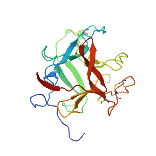
Structure of a Kunitz-type potato cathepsin D inhibitor.

Guo, J.Erskine, P.T.Coker, A.R.Wood, S.P.Cooper, J.B.

(2015) J Struct Biol 192: 554-560

  • DOI: https://doi.org/10.1016/j.jsb.2015.10.020
  • Primary Citation of Related Structures:  
    5DZU

  • PubMed Abstract: 

    Potato cathepsin D inhibitor (PDI) is a glycoprotein of 188 amino acids which can inhibit both the aspartic protease cathepsin D and the serine protease trypsin. Here we report the first X-ray structure of PDI at a resolution of 2.1 Å showing that PDI adopts a β-trefoil fold, which is typical of the Kunitz-family protease inhibitors, with the inhibitory loops protruding from the core. Possible reactive-site loops including one involving a unique disulphide and another involving a protruding 310 helix are identified and docking studies indicate the mode of action of this unusual bi-functional inhibitor.


  • Organizational Affiliation

    Division of Medicine, UCL, Gower Street, London WC1E 6BT, United Kingdom.


Macromolecules
Find similar proteins by:  (by identity cutoff)  |  3D Structure
Entity ID: 1
MoleculeChains Sequence LengthOrganismDetailsImage
Aspartic protease inhibitor 11
A, B
188Solanum tuberosumMutation(s): 0 
UniProt
Find proteins for P16348 (Solanum tuberosum)
Explore P16348 
Go to UniProtKB:  P16348
Entity Groups  
Sequence Clusters30% Identity50% Identity70% Identity90% Identity95% Identity100% Identity
UniProt GroupP16348
Sequence Annotations
Expand
  • Reference Sequence
Experimental Data & Validation

Experimental Data

  • Method: X-RAY DIFFRACTION
  • Resolution: 2.12 Å
  • R-Value Free: 0.270 
  • R-Value Work: 0.191 
  • R-Value Observed: 0.195 
  • Space Group: C 1 2 1
Unit Cell:
Length ( Å )Angle ( ˚ )
a = 75.458α = 90
b = 124.769β = 95.14
c = 37.876γ = 90
Software Package:
Software NamePurpose
REFMACrefinement
XDSdata reduction
Aimlessdata scaling
MOLREPphasing

Structure Validation

View Full Validation Report


Ligand Structure Quality Assessment 


Entry History 

Deposition Data

Revision History  (Full details and data files)

  • Version 1.0: 2015-10-21
    Type: Initial release
  • Version 1.1: 2015-11-18
    Changes: Database references
  • Version 1.2: 2015-11-25
    Changes: Database references
  • Version 1.3: 2018-04-25
    Changes: Data collection
  • Version 1.4: 2020-07-29
    Type: Remediation
    Reason: Carbohydrate remediation
    Changes: Data collection, Derived calculations, Structure summary
  • Version 1.5: 2024-01-10
    Changes: Data collection, Database references, Refinement description, Structure summary