


Crystal structure of 2'AMP bound SaIMPase-II
Dutta, A., Bhattacharyya, S., Das, A.K.To be published.
Experimental Data Snapshot
Entity ID: 1 | |||||
|---|---|---|---|---|---|
| Molecule | Chains | Sequence Length | Organism | Details | Image |
| Inositol monophosphatase | 283 | Staphylococcus aureus | Mutation(s): 0 Gene Names: sas1042 EC: 3.1.3.25 | 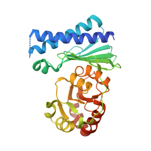 | |
Entity Groups | |||||
| Sequence Clusters | 30% Identity50% Identity70% Identity90% Identity95% Identity100% Identity | ||||
Sequence AnnotationsExpand | |||||
| |||||
| Ligands 3 Unique | |||||
|---|---|---|---|---|---|
| ID | Chains | Name / Formula / InChI Key | 2D Diagram | 3D Interactions | |
| 2AM Query on 2AM | D [auth A], I [auth B] | ADENOSINE-2'-MONOPHOSPHATE C10 H14 N5 O7 P QDFHPFSBQFLLSW-KQYNXXCUSA-N |  | ||
| GOL Query on GOL | F [auth A] | GLYCEROL C3 H8 O3 PEDCQBHIVMGVHV-UHFFFAOYSA-N |  | ||
| CA Query on CA | C [auth A], E [auth A], G [auth B], H [auth B] | CALCIUM ION Ca BHPQYMZQTOCNFJ-UHFFFAOYSA-N |  | ||
| Length ( Å ) | Angle ( ˚ ) |
|---|---|
| a = 49.008 | α = 90 |
| b = 105.687 | β = 101.91 |
| c = 53.313 | γ = 90 |
| Software Name | Purpose |
|---|---|
| SCALA | data scaling |
| MOLREP | phasing |
| REFMAC | refinement |
| PDB_EXTRACT | data extraction |
| XDS | data reduction |
| MOLREP | phasing |
| Funding Organization | Location | Grant Number |
|---|---|---|
| Department of Biotechnology | India | -- |
RCSB PDB (citation) is hosted by
RCSB PDB is a member of the

