5DUO

Crystal structure of native translocator protein 18kDa (TSPO) from Rhodobacter sphaeroides (A139T Mutant) in C2 space group


Experimental Data Snapshot

  • Method: X-RAY DIFFRACTION
  • Resolution: 2.40 Å
  • R-Value Free: 0.244 
  • R-Value Work: 0.189 
  • R-Value Observed: 0.191 

wwPDB Validation   3D Report Full Report


Ligand Structure Quality Assessment 


This is version 1.3 of the entry. See complete history


Literature

Response to Comment on "Crystal structures of translocator protein (TSPO) and mutant mimic of a human polymorphism".

Li, F.Liu, J.Zheng, Y.Garavito, R.M.Ferguson-Miller, S.

(2015) Science 350: 519-519

  • DOI: https://doi.org/10.1126/science.aab2595
  • Primary Citation of Related Structures:  
    5DUO

  • PubMed Abstract: 

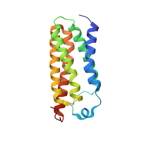
    Wang comments that the diffraction data for the structure of the A139T mutant of translocator protein TSPO from Rhodobacter sphaeroides should be used to 1.65 instead of 1.8 angstroms and that the density interpreted as porphyrin and monoolein is better fitted as polyethylene glycol. Although different practices of data processing exist, in this case they do not substantially influence the final map. Additional data are presented supporting the fit of a porphyrin and monooleins.


  • Organizational Affiliation

    Department of Biochemistry and Molecular Biology, Michigan State University, East Lansing, MI 48824, USA.


Macromolecules
Find similar proteins by:  (by identity cutoff)  |  3D Structure
Entity ID: 1
MoleculeChains Sequence LengthOrganismDetailsImage
Tryptophan-rich sensory protein
A, B, C
157Cereibacter sphaeroidesMutation(s): 1 
Gene Names: tspOcrtK
Membrane Entity: Yes 
UniProt
Find proteins for Q9RFC8 (Cereibacter sphaeroides)
Explore Q9RFC8 
Go to UniProtKB:  Q9RFC8
Entity Groups  
Sequence Clusters30% Identity50% Identity70% Identity90% Identity95% Identity100% Identity
UniProt GroupQ9RFC8
Sequence Annotations
Expand
  • Reference Sequence
Small Molecules
Ligands 3 Unique
IDChains Name / Formula / InChI Key2D Diagram3D Interactions
PP9
Query on PP9

Download Ideal Coordinates CCD File 
K [auth A]PROTOPORPHYRIN IX
C34 H34 N4 O4
FEDYMSUPMFCVOD-UJJXFSCMSA-N
OLC
Query on OLC

Download Ideal Coordinates CCD File 
D [auth A]
E [auth A]
F [auth A]
G [auth A]
H [auth A]
D [auth A],
E [auth A],
F [auth A],
G [auth A],
H [auth A],
L [auth B],
M [auth B],
N [auth B],
O [auth B],
P [auth B],
Q [auth B],
R [auth C],
S [auth C],
T [auth C],
U [auth C]
(2R)-2,3-dihydroxypropyl (9Z)-octadec-9-enoate
C21 H40 O4
RZRNAYUHWVFMIP-GDCKJWNLSA-N
FMT
Query on FMT

Download Ideal Coordinates CCD File 
I [auth A],
J [auth A]
FORMIC ACID
C H2 O2
BDAGIHXWWSANSR-UHFFFAOYSA-N
Experimental Data & Validation

Experimental Data

  • Method: X-RAY DIFFRACTION
  • Resolution: 2.40 Å
  • R-Value Free: 0.244 
  • R-Value Work: 0.189 
  • R-Value Observed: 0.191 
  • Space Group: C 1 2 1
Unit Cell:
Length ( Å )Angle ( ˚ )
a = 58.388α = 90
b = 99.191β = 100.05
c = 95.326γ = 90
Software Package:
Software NamePurpose
PHENIXrefinement
HKL-2000data scaling
PDB_EXTRACTdata extraction
XDSdata reduction
SHARPphasing

Structure Validation

View Full Validation Report


Ligand Structure Quality Assessment 


Entry History & Funding Information

Deposition Data


Funding OrganizationLocationGrant Number
National Institutes of Health/National Institute of General Medical Sciences (NIH/NIGMS)United StatesGM26916

Revision History  (Full details and data files)

  • Version 1.0: 2015-11-11
    Type: Initial release
  • Version 1.1: 2017-09-13
    Changes: Author supporting evidence, Database references, Derived calculations
  • Version 1.2: 2019-12-25
    Changes: Author supporting evidence
  • Version 1.3: 2024-03-06
    Changes: Data collection, Database references