5DTU

Crystal structure of the RNA-helicase Prp28 from Chaetomium thermophilum bound to ADP


Experimental Data Snapshot

  • Method: X-RAY DIFFRACTION
  • Resolution: 3.20 Å
  • R-Value Free: 0.288 
  • R-Value Work: 0.274 
  • R-Value Observed: 0.276 

wwPDB Validation   3D Report Full Report


Ligand Structure Quality Assessment 


This is version 1.2 of the entry. See complete history


Literature

Structural analysis of the spliceosomal RNA helicase Prp28 from the thermophilic eukaryote Chaetomium thermophilum.

Tauchert, M.J.Ficner, R.

(2016) Acta Crystallogr F Struct Biol Commun 72: 409-416

  • DOI: https://doi.org/10.1107/S2053230X16006038
  • Primary Citation of Related Structures:  
    5DTU

  • PubMed Abstract: 

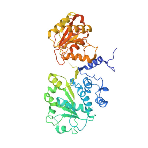
    Prp28 (pre-mRNA-splicing ATP-dependent RNA helicase 28) is a spliceosomal DEAD-box helicase which is involved in two steps of spliceosome assembly. It is required for the formation of commitment complex 2 in an ATP-independent manner as well as for the formation of the pre-catalytic spliceosome, which in contrast is ATP-dependent. During the latter step, Prp28 is crucial for the integration of the U4/U6·U5 tri-snRNP since it displaces the U1 snRNP and allows the U6 snRNP to base-pair with the 5'-splice site. Here, the crystal structure of Prp28 from the thermophilic fungus Chaetomium thermophilum is reported at 3.2 Å resolution and is compared with the available structures of homologues.


  • Organizational Affiliation

    Department of Molecular Structural Biology, Institute for Microbiology and Genetics, GZMB, Georg-August-University Göttingen, Justus-von-Liebig Weg 11, 37077 Göttingen, Germany.


Macromolecules
Find similar proteins by:  (by identity cutoff)  |  3D Structure
Entity ID: 1
MoleculeChains Sequence LengthOrganismDetailsImage
Prp28475Thermochaetoides thermophila DSM 1495Mutation(s): 0 
Gene Names: CTHT_0054430
UniProt
Find proteins for G0SBQ7 (Chaetomium thermophilum (strain DSM 1495 / CBS 144.50 / IMI 039719))
Explore G0SBQ7 
Go to UniProtKB:  G0SBQ7
Entity Groups  
Sequence Clusters30% Identity50% Identity70% Identity90% Identity95% Identity100% Identity
UniProt GroupG0SBQ7
Sequence Annotations
Expand
  • Reference Sequence
Small Molecules
Ligands 2 Unique
IDChains Name / Formula / InChI Key2D Diagram3D Interactions
ADP
Query on ADP

Download Ideal Coordinates CCD File 
B [auth A]ADENOSINE-5'-DIPHOSPHATE
C10 H15 N5 O10 P2
XTWYTFMLZFPYCI-KQYNXXCUSA-N
MG
Query on MG

Download Ideal Coordinates CCD File 
C [auth A]MAGNESIUM ION
Mg
JLVVSXFLKOJNIY-UHFFFAOYSA-N
Experimental Data & Validation

Experimental Data

  • Method: X-RAY DIFFRACTION
  • Resolution: 3.20 Å
  • R-Value Free: 0.288 
  • R-Value Work: 0.274 
  • R-Value Observed: 0.276 
  • Space Group: C 1 2 1
Unit Cell:
Length ( Å )Angle ( ˚ )
a = 161.5α = 90
b = 50.4β = 101.4
c = 65.8γ = 90
Software Package:
Software NamePurpose
PHENIXrefinement
XDSdata reduction
XSCALEdata scaling
PHASERphasing

Structure Validation

View Full Validation Report


Ligand Structure Quality Assessment 


Entry History & Funding Information

Deposition Data


Funding OrganizationLocationGrant Number
German Research FoundationGermanySFB860

Revision History  (Full details and data files)

  • Version 1.0: 2016-05-04
    Type: Initial release
  • Version 1.1: 2016-05-11
    Changes: Database references
  • Version 1.2: 2024-01-10
    Changes: Author supporting evidence, Data collection, Database references, Refinement description