5DBX

Crystal structure of murine SPAK(T243D) in complex with AMPPNP


Experimental Data Snapshot

  • Method: X-RAY DIFFRACTION
  • Resolution: 2.50 Å
  • R-Value Free: 0.246 
  • R-Value Work: 0.206 
  • R-Value Observed: 0.208 

wwPDB Validation   3D Report Full Report


Ligand Structure Quality Assessment 


This is version 1.1 of the entry. See complete history


Literature

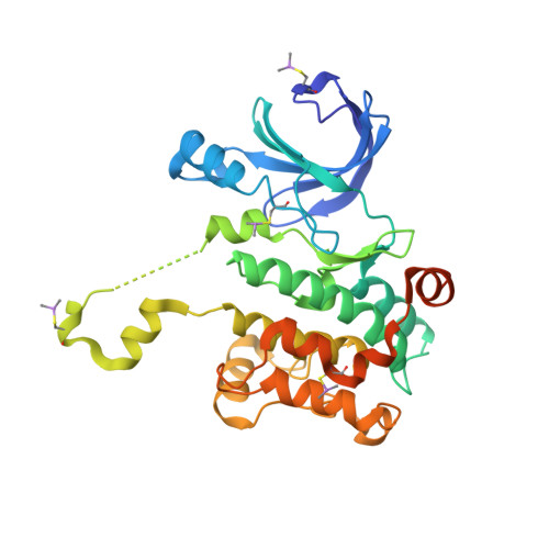
Domain-Swapping Switch Point in Ste20 Protein Kinase SPAK.

Taylor, C.A.Juang, Y.C.Earnest, S.Sengupta, S.Goldsmith, E.J.Cobb, M.H.

(2015) Biochemistry 54: 5063-5071

  • DOI: https://doi.org/10.1021/acs.biochem.5b00593
  • Primary Citation of Related Structures:  
    5D9H, 5DBX

  • PubMed Abstract: 

    The related protein kinases SPAK and OSR1 regulate ion homeostasis in part by phosphorylating cation cotransporter family members. The structure of the kinase domain of OSR1 was determined in the unphosphorylated inactive form and, like some other Ste20 kinases, exhibited a domain-swapped activation loop. To further probe the role of domain swapping in SPAK and OSR1, we have determined the crystal structures of SPAK 63-403 at 3.1 Å and SPAK 63-390 T243D at 2.5 Å resolution. These structures encompass the kinase domain and different portions of the C-terminal tail, the longer without and the shorter with an activating T243D point mutation. The structure of the T243D protein reveals significant conformational differences relative to unphosphorylated SPAK and OSR1 but also has some features of an inactive kinase. Both structures are domain-swapped dimers. Sequences involved in domain swapping were identified and mutated to create a SPAK monomeric mutant with kinase activity, indicating that monomeric forms are active. The monomeric mutant is activated by WNK1 but has reduced activity toward its substrate NKCC2, suggesting regulatory roles for domain swapping. The structure of partially active SPAK T243D is consistent with a multistage activation process in which phosphorylation induces a SPAK conformation that requires further remodeling to build the active structure.


  • Organizational Affiliation

    †Department of Pharmacology and ‡Department of Biophysics, University of Texas Southwestern Medical Center, Dallas, Texas 75390, United States.


Macromolecules
Find similar proteins by:  (by identity cutoff)  |  3D Structure
Entity ID: 1
MoleculeChains Sequence LengthOrganismDetailsImage
STE20/SPS1-related proline-alanine-rich protein kinase
A, B
328Mus musculusMutation(s): 1 
Gene Names: Stk39Spak
EC: 2.7.11.1
UniProt & NIH Common Fund Data Resources
Find proteins for Q9Z1W9 (Mus musculus)
Explore Q9Z1W9 
Go to UniProtKB:  Q9Z1W9
IMPC:  MGI:1858416
Entity Groups  
Sequence Clusters30% Identity50% Identity70% Identity90% Identity95% Identity100% Identity
UniProt GroupQ9Z1W9
Sequence Annotations
Expand
  • Reference Sequence
Small Molecules
Modified Residues  1 Unique
IDChains TypeFormula2D DiagramParent
CAS
Query on CAS
A, B
L-PEPTIDE LINKINGC5 H12 As N O2 SCYS
Experimental Data & Validation

Experimental Data

  • Method: X-RAY DIFFRACTION
  • Resolution: 2.50 Å
  • R-Value Free: 0.246 
  • R-Value Work: 0.206 
  • R-Value Observed: 0.208 
  • Space Group: P 21 21 21
Unit Cell:
Length ( Å )Angle ( ˚ )
a = 66.442α = 90
b = 101.736β = 90
c = 104.077γ = 90
Software Package:
Software NamePurpose
PHENIXrefinement
HKL-2000data reduction
DENZOdata reduction
PHASERphasing

Structure Validation

View Full Validation Report


Ligand Structure Quality Assessment 


Entry History 

Deposition Data

  • Released Date: 2015-09-23 
  • Deposition Author(s): Juang, Y.-C.

Revision History  (Full details and data files)

  • Version 1.0: 2015-09-23
    Type: Initial release
  • Version 1.1: 2023-09-27
    Changes: Data collection, Database references, Derived calculations, Refinement description