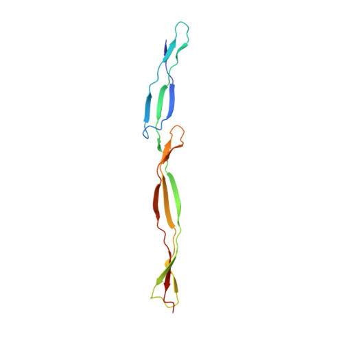
Disorder drives cooperative folding in a multidomain protein.
Gruszka, D.T., Mendonca, C.A., Paci, E., Whelan, F., Hawkhead, J., Potts, J.R., Clarke, J.(2016) Proc Natl Acad Sci U S A 113: 11841-11846
- PubMed: 27698144
- DOI: https://doi.org/10.1073/pnas.1608762113
- Primary Citation of Related Structures:
5DBL - PubMed Abstract:
Many human proteins contain intrinsically disordered regions, and disorder in these proteins can be fundamental to their function-for example, facilitating transient but specific binding, promoting allostery, or allowing efficient posttranslational modification. SasG, a multidomain protein implicated in host colonization and biofilm formation in Staphylococcus aureus, provides another example of how disorder can play an important role. Approximately one-half of the domains in the extracellular repetitive region of SasG are intrinsically unfolded in isolation, but these E domains fold in the context of their neighboring folded G5 domains. We have previously shown that the intrinsic disorder of the E domains mediates long-range cooperativity between nonneighboring G5 domains, allowing SasG to form a long, rod-like, mechanically strong structure. Here, we show that the disorder of the E domains coupled with the remarkable stability of the interdomain interface result in cooperative folding kinetics across long distances. Formation of a small structural nucleus at one end of the molecule results in rapid structure formation over a distance of 10 nm, which is likely to be important for the maintenance of the structural integrity of SasG. Moreover, if this normal folding nucleus is disrupted by mutation, the interdomain interface is sufficiently stable to drive the folding of adjacent E and G5 domains along a parallel folding pathway, thus maintaining cooperative folding.
Organizational Affiliation:
Department of Chemistry, University of Cambridge, Cambridge CB2 1EW, United Kingdom.


