5D29

X-ray structure of human glutamate carboxypeptidase II (GCPII) in complex with a hydroxamate inhibitor JHU241


Experimental Data Snapshot

  • Method: X-RAY DIFFRACTION
  • Resolution: 1.80 Å
  • R-Value Free: 0.205 
  • R-Value Work: 0.174 
  • R-Value Observed: 0.176 

wwPDB Validation   3D Report Full Report


Ligand Structure Quality Assessment 


This is version 2.0 of the entry. See complete history


Literature

Unprecedented Binding Mode of Hydroxamate-Based Inhibitors of Glutamate Carboxypeptidase II: Structural Characterization and Biological Activity.

Novakova, Z.Wozniak, K.Jancarik, A.Rais, R.Wu, Y.Pavlicek, J.Ferraris, D.Havlinova, B.Ptacek, J.Vavra, J.Hin, N.Rojas, C.Majer, P.Slusher, B.S.Tsukamoto, T.Barinka, C.

(2016) J Med Chem 59: 4539-4550

  • DOI: https://doi.org/10.1021/acs.jmedchem.5b01806
  • Primary Citation of Related Structures:  
    5D29, 5ELY

  • PubMed Abstract: 

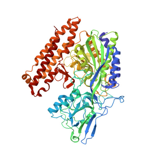
    Inhibition of glutamate carboxypeptidase II (GCPII) is effective in preclinical models of neurological disorders associated with excessive activation of glutamatergic systems. Here we report synthesis, structural characterization, and biological activity of new hydroxamic acid-based inhibitors with nanomolar affinity for human GCPII. Crystal structures of GCPII/hydroxamate complexes revealed an unprecedented binding mode in which the putative P1' glutarate occupies the spacious entrance funnel rather than the conserved glutamate-binding S1' pocket. This unique binding mode provides a mechanistic explanation for the structure-activity relationship data, most notably the lack of enantiospecificity and the tolerance for bulky/hydrophobic functions as substituents of a canonical glutarate moiety. The in vivo pharmacokinetics profile of one of the inhibitors will be presented along with analgesic efficacy data from the rat chronic constrictive injury model of neuropathic pain.


  • Organizational Affiliation

    Institute of Biotechnology, Academy of Sciences of the Czech Republic, BIOCEV , Prumyslova 595, 252 50 Vestec, Czech Republic.


Macromolecules
Find similar proteins by:  (by identity cutoff)  |  3D Structure
Entity ID: 1
MoleculeChains Sequence LengthOrganismDetailsImage
Glutamate carboxypeptidase 2695Homo sapiensMutation(s): 0 
Gene Names: FOLH1FOLHNAALAD1PSMPSMAGIG27
EC: 3.4.17.21
UniProt & NIH Common Fund Data Resources
Find proteins for Q04609 (Homo sapiens)
Explore Q04609 
Go to UniProtKB:  Q04609
PHAROS:  Q04609
GTEx:  ENSG00000086205 
Entity Groups  
Sequence Clusters30% Identity50% Identity70% Identity90% Identity95% Identity100% Identity
UniProt GroupQ04609
Sequence Annotations
Expand
  • Reference Sequence
Oligosaccharides

Help

Entity ID: 2
MoleculeChains Length2D Diagram Glycosylation3D Interactions
2-acetamido-2-deoxy-beta-D-glucopyranose-(1-4)-2-acetamido-2-deoxy-beta-D-glucopyranose
B, C
2N-Glycosylation
Glycosylation Resources
GlyTouCan:  G42666HT
GlyCosmos:  G42666HT
GlyGen:  G42666HT
Entity ID: 3
MoleculeChains Length2D Diagram Glycosylation3D Interactions
beta-D-mannopyranose-(1-3)-beta-D-mannopyranose-(1-4)-2-acetamido-2-deoxy-beta-D-glucopyranose-(1-4)-2-acetamido-2-deoxy-beta-D-glucopyranose
D
4N-Glycosylation
Glycosylation Resources
GlyTouCan:  G31886NL
GlyCosmos:  G31886NL
GlyGen:  G31886NL
Small Molecules
Ligands 6 Unique
IDChains Name / Formula / InChI Key2D Diagram3D Interactions
5Q1
Query on 5Q1

Download Ideal Coordinates CCD File 
N [auth A]4-[(2~{S})-2-carboxy-5-(oxidanylamino)-5-oxidanylidene-pentyl]benzoic acid
C13 H15 N O6
VUDGMYIWOPKVSL-JTQLQIEISA-N
NAG
Query on NAG

Download Ideal Coordinates CCD File 
E [auth A],
F [auth A],
G [auth A],
H [auth A]
2-acetamido-2-deoxy-beta-D-glucopyranose
C8 H15 N O6
OVRNDRQMDRJTHS-FMDGEEDCSA-N
ZN
Query on ZN

Download Ideal Coordinates CCD File 
I [auth A],
J [auth A]
ZINC ION
Zn
PTFCDOFLOPIGGS-UHFFFAOYSA-N
ACT
Query on ACT

Download Ideal Coordinates CCD File 
M [auth A]ACETATE ION
C2 H3 O2
QTBSBXVTEAMEQO-UHFFFAOYSA-M
CA
Query on CA

Download Ideal Coordinates CCD File 
K [auth A]CALCIUM ION
Ca
BHPQYMZQTOCNFJ-UHFFFAOYSA-N
CL
Query on CL

Download Ideal Coordinates CCD File 
L [auth A]CHLORIDE ION
Cl
VEXZGXHMUGYJMC-UHFFFAOYSA-M
Binding Affinity Annotations 
IDSourceBinding Affinity
5Q1 Binding MOAD:  5D29 IC50: 97 (nM) from 1 assay(s)
BindingDB:  5D29 IC50: 97 (nM) from 1 assay(s)
Experimental Data & Validation

Experimental Data

  • Method: X-RAY DIFFRACTION
  • Resolution: 1.80 Å
  • R-Value Free: 0.205 
  • R-Value Work: 0.174 
  • R-Value Observed: 0.176 
  • Space Group: I 2 2 2
Unit Cell:
Length ( Å )Angle ( ˚ )
a = 100.191α = 90
b = 130.459β = 90
c = 157.196γ = 90
Software Package:
Software NamePurpose
REFMACrefinement
PDB_EXTRACTdata extraction
DENZOdata reduction
XSCALEdata scaling
MAR345dtbdata collection

Structure Validation

View Full Validation Report


Ligand Structure Quality Assessment 


Entry History & Funding Information

Deposition Data


Funding OrganizationLocationGrant Number
Helmholtz-Zentrum Berlin and BioStruct-XGermanyN283570

Revision History  (Full details and data files)

  • Version 1.0: 2016-04-27
    Type: Initial release
  • Version 1.1: 2016-06-08
    Changes: Database references
  • Version 2.0: 2020-07-29
    Type: Remediation
    Reason: Carbohydrate remediation
    Changes: Atomic model, Data collection, Derived calculations, Structure summary