5D0E

Crystal Structure of an adenylyl cyclase Ma1120-Cat in complex with GTP and calcium from Mycobacterium avium


Experimental Data Snapshot

  • Method: X-RAY DIFFRACTION
  • Resolution: 1.48 Å
  • R-Value Free: 0.225 
  • R-Value Work: 0.189 
  • R-Value Observed: 0.191 

wwPDB Validation   3D Report Full Report


Ligand Structure Quality Assessment 


This is version 1.3 of the entry. See complete history


Literature

Substrate specificity determinants of class III nucleotidyl cyclases

Bharambe, N.G.Barathy, D.V.Syed, W.Visweswariah, S.S.Cola sigmaf o, M.Misquith, S.Suguna, K.

(2016) FEBS J 283: 3723-3738

  • DOI: https://doi.org/10.1111/febs.13837
  • Primary Citation of Related Structures:  
    5D0E, 5D0G, 5D0H, 5D15

  • PubMed Abstract: 

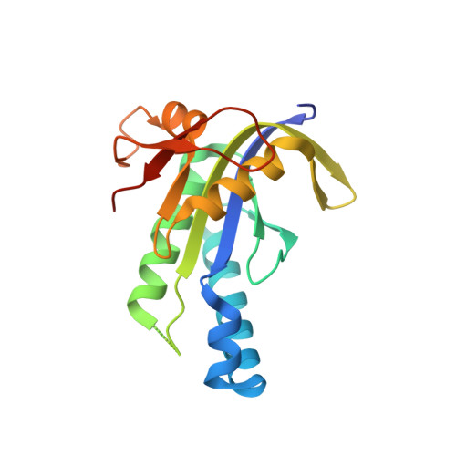
    The two second messengers in signalling, cyclic AMP and cyclic GMP, are produced by adenylyl and guanylyl cyclases respectively. Recognition and discrimination of the substrates ATP and GTP by the nucleotidyl cyclases are vital in these reactions. Various apo-, substrate- or inhibitor-bound forms of adenylyl cyclase (AC) structures from transmembrane and soluble ACs have revealed the catalytic mechanism of ATP cyclization reaction. Previously reported structures of guanylyl cyclases represent ligand-free forms and inactive open states of the enzymes and thus do not provide information regarding the exact mode of substrate binding. The structures we present here of the cyclase homology domain of a class III AC from Mycobacterium avium (Ma1120) and its mutant in complex with ATP and GTP in the presence of calcium ion, provide the structural basis for substrate selection by the nucleotidyl cyclases at the atomic level. Precise nature of the enzyme-substrate interactions, novel modes of substrate binding and the ability of the binding pocket to accommodate diverse conformations of the substrates have been revealed by the present crystallographic analysis. This is the first report to provide structures of both the nucleotide substrates bound to a nucleotidyl cyclase.


  • Organizational Affiliation

    Molecular Biophysics Unit, Indian Institute of Science, Bangalore, India.


Macromolecules
Find similar proteins by:  (by identity cutoff)  |  3D Structure
Entity ID: 1
MoleculeChains Sequence LengthOrganismDetailsImage
Cyclase
A, B
177Mycobacterium avium subsp. avium 10-9275Mutation(s): 0 
Gene Names: O972_06080
UniProt
Find proteins for V7LAR8 (Mycobacterium avium subsp. avium 10-9275)
Explore V7LAR8 
Go to UniProtKB:  V7LAR8
Entity Groups  
Sequence Clusters30% Identity50% Identity70% Identity90% Identity95% Identity100% Identity
UniProt GroupV7LAR8
Sequence Annotations
Expand
  • Reference Sequence
Experimental Data & Validation

Experimental Data

  • Method: X-RAY DIFFRACTION
  • Resolution: 1.48 Å
  • R-Value Free: 0.225 
  • R-Value Work: 0.189 
  • R-Value Observed: 0.191 
  • Space Group: P 1 21 1
Unit Cell:
Length ( Å )Angle ( ˚ )
a = 52.59α = 90
b = 55.71β = 109.74
c = 55.27γ = 90
Software Package:
Software NamePurpose
PHENIXrefinement

Structure Validation

View Full Validation Report


Ligand Structure Quality Assessment 


Entry History 

Deposition Data

Revision History  (Full details and data files)

  • Version 1.0: 2016-08-10
    Type: Initial release
  • Version 1.1: 2016-09-07
    Changes: Database references
  • Version 1.2: 2016-11-02
    Changes: Database references
  • Version 1.3: 2023-11-08
    Changes: Advisory, Data collection, Database references, Derived calculations, Refinement description