5BZA

Crystal structure of CbsA from Thermotoga neapolitana


Experimental Data Snapshot

  • Method: X-RAY DIFFRACTION
  • Resolution: 2.00 Å
  • R-Value Free: 0.276 
  • R-Value Work: 0.238 
  • R-Value Observed: 0.238 

wwPDB Validation   3D Report Full Report


This is version 1.1 of the entry. See complete history


Literature

Crystal structure of beta-N-acetylglucosaminidase CbsA from Thermotoga neapolitana

Kim, J.S.Yoon, B.Y.Ahn, J.Cha, J.Ha, N.C.

(2015) Biochem Biophys Res Commun 464: 869-874

  • DOI: https://doi.org/10.1016/j.bbrc.2015.07.053
  • Primary Citation of Related Structures:  
    5BZA, 5C0Q

  • PubMed Abstract: 

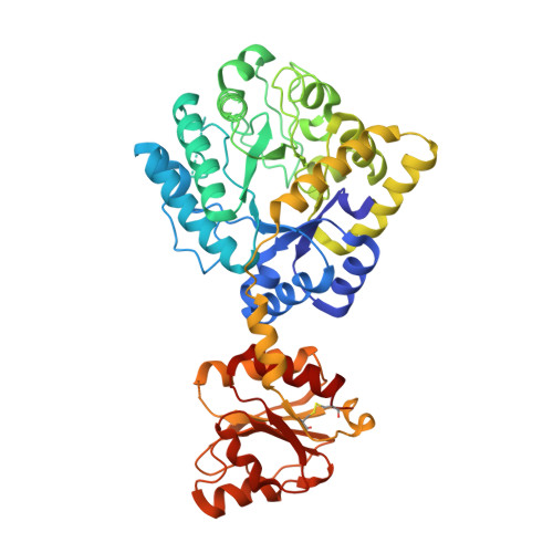
    CbsA from the thermophilic marine bacteria Thermotoga neapolitana is a chitinolyitc enzyme that can cleave a glycosidic bond of the polymer N-acetylglucosamine at the non-reducing end. This enzyme has particularly high activity on di-N-acetylchitobiose. CbsA consists of a family of 3 glycoside hydrolase (GH3)-type catalytic domains and a unique C-terminal domain. The C-terminal domain distinguishes CbsA from other GH3-type enzymes. Sequence analyses have suggested that CbsA has the Asp-His dyad as a general acid/base with the NagZ of Bacillus subtilis and the Salmonella enterica serovar Typhimurium. Here, we determined the crystal structure of CbsA from T. neapolitana at a resolution of 2.0 Å using the Zn-SAD method, revealing a unique homodimeric assembly facilitated by the C-terminal domains in the dimer. We observed that CbsA is strongly inhibited by ZnCl2, and two zinc ions were consistently bound in the active site. Our results can explain the zinc ion's inhibition mechanism in the subfamily of GH3 enzymes, and provide information on the structural diversity and substrate specificity of this hydrolase family.


  • Organizational Affiliation

    Department of Agricultural Biotechnology, Center for Food Safety and Toxicology, Center for Food and Bioconvergence, Research Institute for Agricultural and Life Sciences, Seoul National University, Seoul, Republic of Korea.


Macromolecules
Find similar proteins by:  (by identity cutoff)  |  3D Structure
Entity ID: 1
MoleculeChains Sequence LengthOrganismDetailsImage
Beta-N-acetylhexosaminidase
A, B, C, D
467Thermotoga neapolitanaMutation(s): 0 
Gene Names: cbsA
EC: 3.2.1.52
UniProt
Find proteins for Q9AG27 (Thermotoga neapolitana)
Explore Q9AG27 
Go to UniProtKB:  Q9AG27
Entity Groups  
Sequence Clusters30% Identity50% Identity70% Identity90% Identity95% Identity100% Identity
UniProt GroupQ9AG27
Sequence Annotations
Expand
  • Reference Sequence
Small Molecules
Ligands 1 Unique
IDChains Name / Formula / InChI Key2D Diagram3D Interactions
CD
Query on CD

Download Ideal Coordinates CCD File 
AA [auth C]
BA [auth C]
CA [auth C]
DA [auth C]
E [auth A]
AA [auth C],
BA [auth C],
CA [auth C],
DA [auth C],
E [auth A],
EA [auth D],
F [auth A],
FA [auth D],
G [auth A],
GA [auth D],
H [auth A],
HA [auth D],
I [auth A],
IA [auth D],
J [auth A],
JA [auth D],
K [auth A],
KA [auth D],
L [auth A],
M [auth B],
N [auth B],
O [auth B],
P [auth B],
Q [auth B],
R [auth B],
S [auth B],
T [auth B],
U [auth B],
V [auth C],
W [auth C],
X [auth C],
Y [auth C],
Z [auth C]
CADMIUM ION
Cd
WLZRMCYVCSSEQC-UHFFFAOYSA-N
Experimental Data & Validation

Experimental Data

  • Method: X-RAY DIFFRACTION
  • Resolution: 2.00 Å
  • R-Value Free: 0.276 
  • R-Value Work: 0.238 
  • R-Value Observed: 0.238 
  • Space Group: H 3 2
Unit Cell:
Length ( Å )Angle ( ˚ )
a = 158.965α = 90
b = 158.965β = 90
c = 517.242γ = 120
Software Package:
Software NamePurpose
PHENIXrefinement
HKL-2000data reduction
HKL-2000data scaling
PHENIXphasing

Structure Validation

View Full Validation Report


Entry History 

Deposition Data

Revision History  (Full details and data files)

  • Version 1.0: 2015-09-16
    Type: Initial release
  • Version 1.1: 2020-02-19
    Changes: Data collection, Database references, Derived calculations