5BXH

Crystal structure of pentameric KCTD9 BTB domain


Experimental Data Snapshot

  • Method: X-RAY DIFFRACTION
  • Resolution: 2.76 Å
  • R-Value Free: 0.280 
  • R-Value Work: 0.234 
  • R-Value Observed: 0.237 

wwPDB Validation   3D Report Full Report


This is version 1.3 of the entry. See complete history


Literature

Structural Insights into KCTD Protein Assembly and Cullin3 Recognition.

Ji, A.X.Chu, A.Nielsen, T.K.Benlekbir, S.Rubinstein, J.L.Prive, G.G.

(2016) J Mol Biol 428: 92-107

  • DOI: https://doi.org/10.1016/j.jmb.2015.08.019
  • Primary Citation of Related Structures:  
    5BXB, 5BXD, 5BXH

  • PubMed Abstract: 

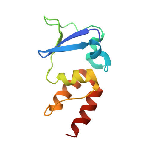
    Cullin3 (Cul3)-based ubiquitin E3 ligase complexes catalyze the transfer of ubiquitin from an E2 enzyme to target substrate proteins. In these assemblies, the C-terminal region of Cul3 binds Rbx1/E2-ubiquitin, while the N-terminal region interacts with various BTB (bric-à-brac, tramtrack, broad complex) domain proteins that serve as substrate adaptors. Previous crystal structures of the homodimeric BTB proteins KLHL3, KLHL11 and SPOP in complex with the N-terminal domain of Cul3 revealed the features required for Cul3 recognition in these proteins. A second class of BTB-domain-containing proteins, the KCTD proteins, is also Cul3 substrate adaptors, but these do not share many of the previously identified determinants for Cul3 binding. We report the pentameric crystal structures of the KCTD1 and KCTD9 BTB domains and identify plasticity in the KCTD1 rings. We find that the KCTD proteins 5, 6, 9 and 17 bind to Cul3 with high affinity, while the KCTD proteins 1 and 16 do not have detectable binding. Finally, we confirm the 5:5 assembly of KCTD9/Cul3 complexes by cryo-electron microscopy and provide a molecular rationale for BTB-mediated Cul3 binding specificity in the KCTD family.


  • Organizational Affiliation

    Department of Biochemistry, University of Toronto, 1 King's College Circle, Toronto, ON, M5S 1A8, Canada.


Macromolecules
Find similar proteins by:  (by identity cutoff)  |  3D Structure
Entity ID: 1
MoleculeChains Sequence LengthOrganismDetailsImage
BTB/POZ domain-containing protein KCTD9
A, B, C, D, E
106Homo sapiensMutation(s): 0 
Gene Names: KCTD9
UniProt & NIH Common Fund Data Resources
Find proteins for Q7L273 (Homo sapiens)
Explore Q7L273 
Go to UniProtKB:  Q7L273
PHAROS:  Q7L273
GTEx:  ENSG00000104756 
Entity Groups  
Sequence Clusters30% Identity50% Identity70% Identity90% Identity95% Identity100% Identity
UniProt GroupQ7L273
Sequence Annotations
Expand
  • Reference Sequence
Experimental Data & Validation

Experimental Data

  • Method: X-RAY DIFFRACTION
  • Resolution: 2.76 Å
  • R-Value Free: 0.280 
  • R-Value Work: 0.234 
  • R-Value Observed: 0.237 
  • Space Group: P 31 2 1
Unit Cell:
Length ( Å )Angle ( ˚ )
a = 105.396α = 90
b = 105.396β = 90
c = 97.316γ = 120
Software Package:
Software NamePurpose
PHENIXrefinement
PHENIXphasing
XDSdata reduction
Aimlessdata scaling

Structure Validation

View Full Validation Report


Entry History 

Deposition Data

Revision History  (Full details and data files)

  • Version 1.0: 2015-09-02
    Type: Initial release
  • Version 1.1: 2015-09-23
    Changes: Data collection, Database references
  • Version 1.2: 2016-02-17
    Changes: Database references
  • Version 1.3: 2023-09-27
    Changes: Data collection, Database references, Derived calculations, Refinement description