


EPSP synthase from Acinetobacter baumannii
Sutton, K.A., Schultz, L.W., Russo, T.A., Breen, J., Umland, T.C.(2016) Acta Crystallogr F Struct Biol Commun
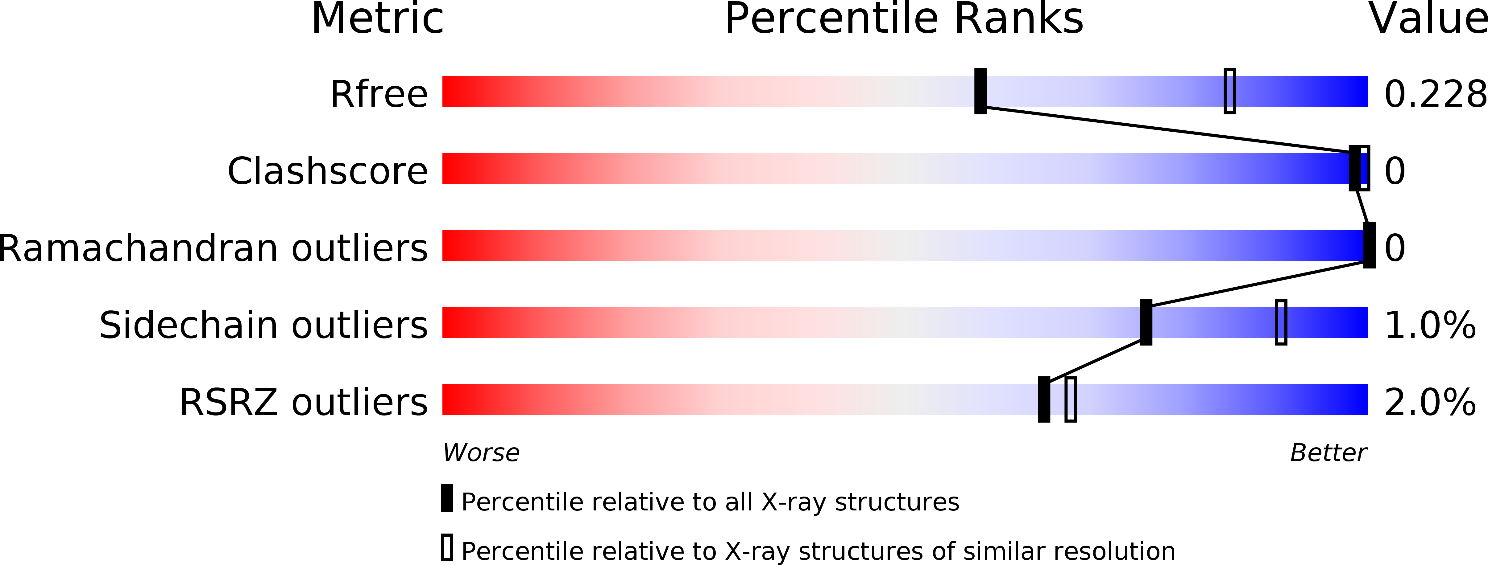
Experimental Data Snapshot
wwPDB Validation 3D Report Full Report
(2016) Acta Crystallogr F Struct Biol Commun
Entity ID: 1 | |||||
|---|---|---|---|---|---|
| Molecule | Chains | Sequence Length | Organism | Details | Image |
| 3-phosphoshikimate 1-carboxyvinyltransferase | 445 | Acinetobacter baumannii ATCC 17978 | Mutation(s): 0 Gene Names: A1S_2276 EC: 2.5.1.19 | 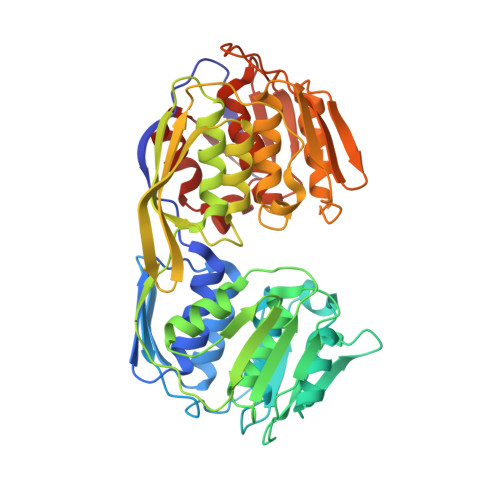 | |
Entity Groups | |||||
| Sequence Clusters | 30% Identity50% Identity70% Identity90% Identity95% Identity100% Identity | ||||
Sequence AnnotationsExpand | |||||
| |||||
| Length ( Å ) | Angle ( ˚ ) |
|---|---|
| a = 73.807 | α = 90 |
| b = 103.896 | β = 90 |
| c = 113.155 | γ = 90 |
| Software Name | Purpose |
|---|---|
| HKL-2000 | data collection |
| HKL-2000 | data scaling |
| PHENIX | refinement |
| PDB_EXTRACT | data extraction |
| PHENIX | phasing |
RCSB PDB (citation) is hosted by
RCSB PDB is a member of the

