5B41

Crystal structure of VDR-LBD complexed with 2-methylidene-19-nor-1a,25-dihydroxyvitamin D3


Experimental Data Snapshot

  • Method: X-RAY DIFFRACTION
  • Resolution: 1.89 Å
  • R-Value Free: 0.234 
  • R-Value Work: 0.187 
  • R-Value Observed: 0.189 

wwPDB Validation   3D Report Full Report


Ligand Structure Quality Assessment 


This is version 1.2 of the entry. See complete history


Literature

Apo- and Antagonist-Binding Structures of Vitamin D Receptor Ligand-Binding Domain Revealed by Hybrid Approach Combining Small-Angle X-ray Scattering and Molecular Dynamics

Anami, Y.Shimizu, N.Ekimoto, T.Egawa, D.Itoh, T.Ikeguchi, M.Yamamoto, K.

(2016) J Med Chem 59: 7888-7900

  • DOI: https://doi.org/10.1021/acs.jmedchem.6b00682
  • Primary Citation of Related Structures:  
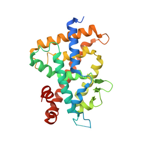
    5B41

  • PubMed Abstract: 

    Vitamin D receptor (VDR) controls the expression of numerous genes through the conformational change caused by binding 1α,25-dihydroxyvitamin D3. Helix 12 in the ligand-binding domain (LBD) is key to regulating VDR activation. The structures of apo VDR-LBD and the VDR-LBD/antagonist complex are unclear. Here, we reveal their unprecedented structures in solution using a hybrid method combining small-angle X-ray scattering and molecular dynamics simulations. In apo rat VDR-LBD, helix 12 is partially unraveled, and it is positioned around the canonical active position and fluctuates. Helix 11 greatly bends toward the outside at Q396, creating a kink. In the rat VDR-LBD/antagonist complex, helix 12 does not generate the activation function 2 surface, and loop 11-12 is remarkably flexible compared to that in the apo rat VDR-LBD. On the basis of these structural insights, we propose a "folding-door model" to describe the mechanism of agonism/antagonism of VDR-LBD.


  • Organizational Affiliation

    Laboratory of Drug Design and Medicinal Chemistry, Showa Pharmaceutical University , 3-3165 Higashi-Tamagawagakuen, Machida, Tokyo 194-8543, Japan.


Macromolecules
Find similar proteins by:  (by identity cutoff)  |  3D Structure
Entity ID: 1
MoleculeChains Sequence LengthOrganismDetailsImage
Vitamin D3 receptor271Rattus norvegicusMutation(s): 0 
Gene Names: VdrNr1i1
UniProt
Find proteins for P13053 (Rattus norvegicus)
Explore P13053 
Go to UniProtKB:  P13053
Entity Groups  
Sequence Clusters30% Identity50% Identity70% Identity90% Identity95% Identity100% Identity
UniProt GroupP13053
Sequence Annotations
Expand
  • Reference Sequence

Find similar proteins by:  Sequence   |   3D Structure  

Entity ID: 2
MoleculeChains Sequence LengthOrganismDetailsImage
Mediator of RNA polymerase II transcription subunit 1B [auth C]13Homo sapiensMutation(s): 0 
UniProt & NIH Common Fund Data Resources
Find proteins for Q15648 (Homo sapiens)
Explore Q15648 
Go to UniProtKB:  Q15648
PHAROS:  Q15648
GTEx:  ENSG00000125686 
Entity Groups  
Sequence Clusters30% Identity50% Identity70% Identity90% Identity95% Identity100% Identity
UniProt GroupQ15648
Sequence Annotations
Expand
  • Reference Sequence
Small Molecules
Ligands 1 Unique
IDChains Name / Formula / InChI Key2D Diagram3D Interactions
YSV
Query on YSV

Download Ideal Coordinates CCD File 
C [auth A](1R,3R)-5-(2-((1R,3aS,7aR,E)-1-((R)-6-hydroxy-6-methylheptan-2-yl)-7a-methyloctahydro-4H-inden-4-ylidene)ethylidene)-2- methylenecyclohexane-1,3-diol
C27 H44 O3
UHMPCVGLSKFXHR-JUTHDLPNSA-N
Binding Affinity Annotations 
IDSourceBinding Affinity
YSV BindingDB:  5B41 Ki: 0.03 (nM) from 1 assay(s)
Experimental Data & Validation

Experimental Data

  • Method: X-RAY DIFFRACTION
  • Resolution: 1.89 Å
  • R-Value Free: 0.234 
  • R-Value Work: 0.187 
  • R-Value Observed: 0.189 
  • Space Group: C 1 2 1
Unit Cell:
Length ( Å )Angle ( ˚ )
a = 154.17α = 90
b = 41.75β = 96.02
c = 41.77γ = 90
Software Package:
Software NamePurpose
REFMACrefinement

Structure Validation

View Full Validation Report


Ligand Structure Quality Assessment 


Entry History 

Deposition Data

Revision History  (Full details and data files)

  • Version 1.0: 2016-12-07
    Type: Initial release
  • Version 1.1: 2020-02-26
    Changes: Data collection, Database references
  • Version 1.2: 2023-11-08
    Changes: Data collection, Database references, Derived calculations, Refinement description, Structure summary