5B2H

Crystal structure of HA33 from Clostridium botulinum serotype C strain Yoichi


Experimental Data Snapshot

  • Method: X-RAY DIFFRACTION
  • Resolution: 2.20 Å
  • R-Value Free: 0.213 
  • R-Value Work: 0.184 
  • R-Value Observed: 0.186 

wwPDB Validation   3D Report Full Report


This is version 1.3 of the entry. See complete history


Literature

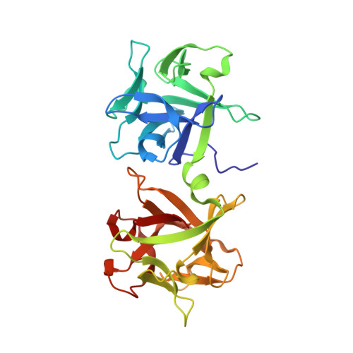
Conformational divergence in the HA-33/HA-17 trimer of serotype C and D botulinum toxin complex

Sagane, Y.Hayashi, S.Akiyama, T.Matsumoto, T.Hasegawa, K.Yamano, A.Suzuki, T.Niwa, K.Watanabe, T.Yajima, S.

(2016) Biochem Biophys Res Commun 476: 280-285

  • DOI: https://doi.org/10.1016/j.bbrc.2016.05.113
  • Primary Citation of Related Structures:  
    5B2H

  • PubMed Abstract: 

    Clostridium botulinum produces a large toxin complex (L-TC) comprising botulinum neurotoxin associated with auxiliary nontoxic proteins. A complex of 33- and 17-kDa hemagglutinins (an HA-33/HA-17 trimer) enhances L-TC transport across the intestinal epithelial cell layer via binding HA-33 to a sugar on the cell surface. At least two subtypes of serotype C/D HA-33 exhibit differing preferences for the sugars sialic acid and galactose. Here, we compared the three-dimensional structures of the galactose-binding HA-33 and HA-33/HA-17 trimers produced by the C-Yoichi strain. Comparisons of serotype C/D HA-33 sequences reveal a variable region with relatively low sequence similarity across the C. botulinum strains; the variability of this region may influence the manner of sugar-recognition by HA-33. Crystal structures of sialic acid- and galactose-binding HA-33 are broadly similar in appearance. However, small-angle X-ray scattering revealed distinct solution structures for HA-33/HA-17 trimers. A structural change in the C-terminal variable region of HA-33 might cause a dramatic shift in the conformation and sugar-recognition mode of HA-33/HA-17 trimer.


  • Organizational Affiliation

    Department of Food and Cosmetic Science, Faculty of Bioindustry, Tokyo University of Agriculture, 196 Yasaka, Abashiri 099-2493, Japan. Electronic address: y3sagane@bioindustry.nodai.ac.jp.


Macromolecules
Find similar proteins by:  (by identity cutoff)  |  3D Structure
Entity ID: 1
MoleculeChains Sequence LengthOrganismDetailsImage
HA-33
A, B
283Clostridium botulinumMutation(s): 0 
Gene Names: ha-33
UniProt
Find proteins for Q93HT5 (Clostridium botulinum)
Explore Q93HT5 
Go to UniProtKB:  Q93HT5
Entity Groups  
Sequence Clusters30% Identity50% Identity70% Identity90% Identity95% Identity100% Identity
UniProt GroupQ93HT5
Sequence Annotations
Expand
  • Reference Sequence
Small Molecules
Ligands 1 Unique
IDChains Name / Formula / InChI Key2D Diagram3D Interactions
PGE
Query on PGE

Download Ideal Coordinates CCD File 
C [auth B]TRIETHYLENE GLYCOL
C6 H14 O4
ZIBGPFATKBEMQZ-UHFFFAOYSA-N
Experimental Data & Validation

Experimental Data

  • Method: X-RAY DIFFRACTION
  • Resolution: 2.20 Å
  • R-Value Free: 0.213 
  • R-Value Work: 0.184 
  • R-Value Observed: 0.186 
  • Space Group: H 3
Unit Cell:
Length ( Å )Angle ( ˚ )
a = 142.517α = 90
b = 142.517β = 90
c = 126.794γ = 120
Software Package:
Software NamePurpose
HKL-2000data collection
HKL-2000data scaling
REFMACrefinement
PDB_EXTRACTdata extraction
Cootmodel building

Structure Validation

View Full Validation Report


Entry History 

Deposition Data

Revision History  (Full details and data files)

  • Version 1.0: 2016-06-15
    Type: Initial release
  • Version 1.1: 2016-07-20
    Changes: Database references
  • Version 1.2: 2020-02-26
    Changes: Data collection, Database references, Derived calculations
  • Version 1.3: 2023-11-08
    Changes: Data collection, Database references, Refinement description