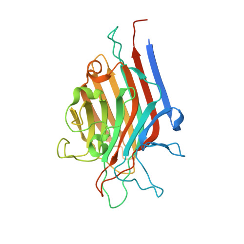
5AVA

Crystal structure of PHA-E lectin in complex with bisected glycan


Experimental Data Snapshot

  • Method: X-RAY DIFFRACTION
  • Resolution: 3.00 Å
  • R-Value Free: 0.274 
  • R-Value Work: 0.229 
  • R-Value Observed: 0.231 

wwPDB Validation   3D Report Full Report


This is version 2.1 of the entry. See complete history


Literature

Atomic visualization of a flipped-back conformation of bisected glycans bound to specific lectins

Nagae, M.Kanagawa, M.Morita-Matsumoto, K.Hanashima, S.Kizuka, Y.Taniguchi, N.Yamaguchi, Y.

(2016) Sci Rep 6: 22973-22973

  • DOI: https://doi.org/10.1038/srep22973
  • Primary Citation of Related Structures:  
    5AV7, 5AVA

  • PubMed Abstract: 

    Glycans normally exist as a dynamic equilibrium of several conformations. A fundamental question concerns how such molecules bind lectins despite disadvantageous entropic loss upon binding. Bisected glycan, a glycan possessing bisecting N-acetylglucosamine (GlcNAc), is potentially a good model for investigating conformational dynamics and glycan-lectin interactions, owing to the unique ability of this sugar residue to alter conformer populations and thus modulate the biological activities. Here we analyzed bisected glycan in complex with two unrelated lectins, Calsepa and PHA-E. The crystal structures of the two complexes show a conspicuous flipped back glycan structure (designated 'back-fold' conformation), and solution NMR analysis also provides evidence of 'back-fold' glycan structure. Indeed, statistical conformational analysis of available bisected and non-bisected glycan structures suggests that bisecting GlcNAc restricts the conformations of branched structures. Restriction of glycan flexibility by certain sugar residues may be more common than previously thought and impinges on the mechanism of glycoform-dependent biological functions.


  • Organizational Affiliation

    Structural Glycobiology Team, 2-1 Hirosawa, Wako, Saitama 351-0198, Japan.


Macromolecules
Find similar proteins by:  (by identity cutoff)  |  3D Structure
Entity ID: 1
MoleculeChains Sequence LengthOrganismDetailsImage
Erythroagglutinin
A, B, C, D, E
A, B, C, D, E, F, G, H
275Phaseolus vulgarisMutation(s): 0 
UniProt
Find proteins for P05088 (Phaseolus vulgaris)
Explore P05088 
Go to UniProtKB:  P05088
Entity Groups  
Sequence Clusters30% Identity50% Identity70% Identity90% Identity95% Identity100% Identity
UniProt GroupP05088
Sequence Annotations
Expand
  • Reference Sequence
Oligosaccharides

Help

Entity ID: 2
MoleculeChains Length2D Diagram Glycosylation3D Interactions
beta-D-galactopyranose-(1-4)-2-acetamido-2-deoxy-beta-D-glucopyranose-(1-2)-alpha-D-mannopyranose-(1-6)-[2-acetamido-2-deoxy-beta-D-glucopyranose-(1-2)-alpha-D-mannopyranose-(1-3)][2-acetamido-2-deoxy-beta-D-glucopyranose-(1-4)]beta-D-mannopyranose-(1-4)-2-acetamido-2-deoxy-beta-D-glucopyranose
I, J, K, L, M
I, J, K, L, M, N, O, P
8N/A
Glycosylation Resources
GlyTouCan:  G67881EE
GlyCosmos:  G67881EE
GlyGen:  G67881EE
Small Molecules
Ligands 2 Unique
IDChains Name / Formula / InChI Key2D Diagram3D Interactions
MN
Query on MN

Download Ideal Coordinates CCD File 
AA [auth F]
CA [auth G]
EA [auth H]
Q [auth A]
S [auth B]
AA [auth F],
CA [auth G],
EA [auth H],
Q [auth A],
S [auth B],
U [auth C],
W [auth D],
Y [auth E]
MANGANESE (II) ION
Mn
WAEMQWOKJMHJLA-UHFFFAOYSA-N
CA
Query on CA

Download Ideal Coordinates CCD File 
BA [auth F]
DA [auth G]
FA [auth H]
R [auth A]
T [auth B]
BA [auth F],
DA [auth G],
FA [auth H],
R [auth A],
T [auth B],
V [auth C],
X [auth D],
Z [auth E]
CALCIUM ION
Ca
BHPQYMZQTOCNFJ-UHFFFAOYSA-N
Experimental Data & Validation

Experimental Data

  • Method: X-RAY DIFFRACTION
  • Resolution: 3.00 Å
  • R-Value Free: 0.274 
  • R-Value Work: 0.229 
  • R-Value Observed: 0.231 
  • Space Group: P 1 21 1
Unit Cell:
Length ( Å )Angle ( ˚ )
a = 95.21α = 90
b = 122.838β = 90.66
c = 97.664γ = 90
Software Package:
Software NamePurpose
REFMACrefinement
HKL-2000data reduction
HKL-2000data scaling
MOLREPphasing

Structure Validation

View Full Validation Report


Entry History & Funding Information

Deposition Data


Funding OrganizationLocationGrant Number
MEXTJapan15K18496, 26110724

Revision History  (Full details and data files)

  • Version 1.0: 2016-04-27
    Type: Initial release
  • Version 1.1: 2020-02-19
    Changes: Data collection, Derived calculations
  • Version 2.0: 2020-07-29
    Type: Remediation
    Reason: Carbohydrate remediation
    Changes: Advisory, Atomic model, Data collection, Derived calculations, Structure summary
  • Version 2.1: 2023-11-08
    Changes: Data collection, Database references, Derived calculations, Refinement description, Structure summary