


Crystal Structure of Wild Type Pneumolysin
Van Pee, K., Yildiz, O.To be published.
Experimental Data Snapshot
wwPDB Validation 3D Report Full Report

Entity ID: 1 | |||||
|---|---|---|---|---|---|
| Molecule | Chains | Sequence Length | Organism | Details | Image |
| PNEUMOLYSIN | 474 | Streptococcus pneumoniae | Mutation(s): 0 Membrane Entity: Yes | 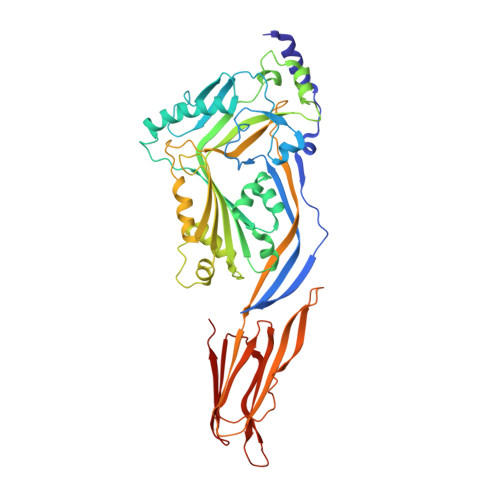 | |
UniProt | |||||
Find proteins for Q7ZAK5 (Streptococcus pneumoniae (strain ATCC BAA-255 / R6)) Explore Q7ZAK5 Go to UniProtKB: Q7ZAK5 | |||||
Entity Groups | |||||
| Sequence Clusters | 30% Identity50% Identity70% Identity90% Identity95% Identity100% Identity | ||||
| UniProt Group | Q7ZAK5 | ||||
Sequence AnnotationsExpand | |||||
| |||||
| Length ( Å ) | Angle ( ˚ ) |
|---|---|
| a = 24.28 | α = 90 |
| b = 113.26 | β = 90 |
| c = 156.67 | γ = 90 |
| Software Name | Purpose |
|---|---|
| PHENIX | refinement |
| XDS | data reduction |
| XSCALE | data scaling |
| PHASER | phasing |
RCSB PDB (citation) is hosted by
RCSB PDB is a member of the

