5AJ8

Tubulin Binding Cofactor C from Leishmania major


Experimental Data Snapshot

  • Method: X-RAY DIFFRACTION
  • Resolution: 2.20 Å
  • R-Value Free: 0.197 
  • R-Value Work: 0.155 
  • R-Value Observed: 0.158 

wwPDB Validation   3D Report Full Report


This is version 1.2 of the entry. See complete history


Literature

Crystal Structure of the C-Terminal Domain of Tubulin-Binding Cofactor C from Leishmania Major.

Barrack, K.L.Fyfe, P.K.Finney, A.J.Hunter, W.N.

(2015) Mol Biochem Parasitol 201: 26

  • DOI: https://doi.org/10.1016/j.molbiopara.2015.05.003
  • Primary Citation of Related Structures:  
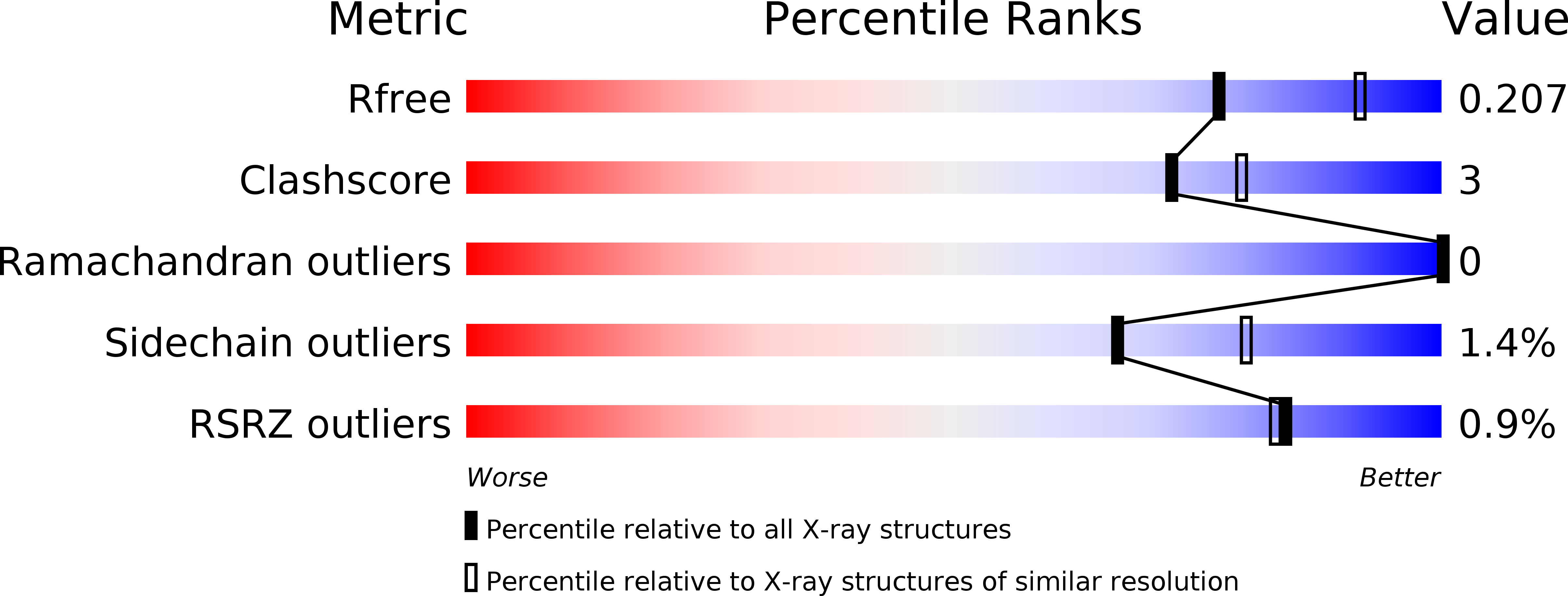
    5AJ8

  • PubMed Abstract: 

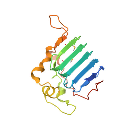
    Tubulin-binding cofactor C stimulates GTPase activity and contributes to the release of the heterodimeric α/β-tubulin from a super-complex of tubulin monomers and two ancillary cofactors. We have determined the 2.2 Å resolution crystal structure of the C-terminal domain of tubulin-binding cofactor C from Leishmania major based on single wavelength anomalous dispersion measurements targeting a selenomethionine derivative. Although previously predicted to consist of two domains the structure is best described as a single domain dominated by a right-handed β-helix of five turns that form a triangular prism. One face of the prism is covered by the C-terminal residues leaving another face solvent exposed. Comparisons with an orthologous human GTPase activating protein match key residues involved in binding nucleotide and identify the face of the β-helix fold likely involved in interacting with the β-tubulin:GTP complex.


  • Organizational Affiliation

    Division of Biological Chemistry & Drug Discovery, College of Life Sciences, University of Dundee, Dundee DD1 5EH, Scotland, UK.


Macromolecules
Find similar proteins by:  (by identity cutoff)  |  3D Structure
Entity ID: 1
MoleculeChains Sequence LengthOrganismDetailsImage
TUBULIN BINDING COFACTOR C
A, B
187Leishmania majorMutation(s): 1 
UniProt
Find proteins for Q4Q1A3 (Leishmania major)
Explore Q4Q1A3 
Go to UniProtKB:  Q4Q1A3
Entity Groups  
Sequence Clusters30% Identity50% Identity70% Identity90% Identity95% Identity100% Identity
UniProt GroupQ4Q1A3
Sequence Annotations
Expand
  • Reference Sequence
Small Molecules
Modified Residues  1 Unique
IDChains TypeFormula2D DiagramParent
MSE
Query on MSE
A, B
L-PEPTIDE LINKINGC5 H11 N O2 SeMET
Experimental Data & Validation

Experimental Data

  • Method: X-RAY DIFFRACTION
  • Resolution: 2.20 Å
  • R-Value Free: 0.197 
  • R-Value Work: 0.155 
  • R-Value Observed: 0.158 
  • Space Group: P 1 21 1
Unit Cell:
Length ( Å )Angle ( ˚ )
a = 37.644α = 90
b = 93.236β = 108.38
c = 48.277γ = 90
Software Package:
Software NamePurpose
REFMACrefinement
XDSdata reduction
Aimlessdata scaling
PHENIXphasing

Structure Validation

View Full Validation Report


Entry History 

Deposition Data

Revision History  (Full details and data files)

  • Version 1.0: 2015-04-15
    Type: Initial release
  • Version 1.1: 2015-06-03
    Changes: Database references
  • Version 1.2: 2015-08-26
    Changes: Database references