5AHV

Cryo-EM structure of helical ANTH and ENTH tubules on PI(4,5)P2-containing membranes


Experimental Data Snapshot

  • Method: ELECTRON MICROSCOPY
  • Resolution: 13.6 Å
  • Aggregation State: FILAMENT 
  • Reconstruction Method: SINGLE PARTICLE 

wwPDB Validation   3D Report Full Report


This is version 2.2 of the entry. See complete history


Literature

An Organized Co-Assembly of Clathrin Adaptors is Essential for Endocytosis.

Skruzny, M.Desfosses, A.Prinz, S.Dodonova, S.O.Gieras, A.Uetrecht, C.Jakobi, A.J.Abella, M.Hagen, W.J.H.Schulz, J.Meijers, R.Rybin, V.Briggs, J.A.G.Sachse, C.Kaksonen, M.

(2015) Dev Cell 33: 150

  • DOI: https://doi.org/10.1016/j.devcel.2015.02.023
  • Primary Citation of Related Structures:  
    5AHV

  • PubMed Abstract: 

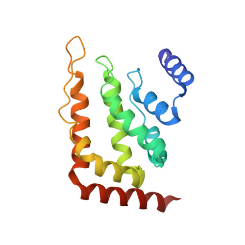
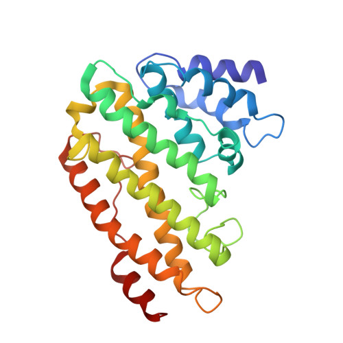
    Clathrin-mediated endocytosis, the main trafficking route from the plasma membrane to the cytoplasm, is critical to many fundamental cellular processes. Clathrin, coupled to the membrane by adaptor proteins, is thought to play a major structural role in endocytosis by self-assembling into a cage-like lattice around the forming vesicle. Although clathrin adaptors are essential for endocytosis, little is known about their structural role in this process. Here we show that the membrane-binding domains of two conserved clathrin adaptors, Sla2 and Ent1, co-assemble in a PI(4,5)P2-dependent manner to form organized lattices on membranes. We determined the structure of the co-assembled lattice by electron cryo-microscopy and designed mutations that specifically impair the lattice formation in vitro. We show that these mutations block endocytosis in vivo. We suggest that clathrin adaptors not only link the polymerized clathrin to the membrane but also form an oligomeric structure, which is essential for membrane remodeling during endocytosis.


  • Organizational Affiliation

    Cell Biology and Biophysics Unit, European Molecular Biology Laboratory (EMBL), 69117 Heidelberg, Germany.


Macromolecules
Find similar proteins by:  (by identity cutoff)  |  3D Structure
Entity ID: 1
MoleculeChains Sequence LengthOrganismDetailsImage
ENTH DOMAIN OF EPSIN ENT1A [auth E]154Saccharomyces cerevisiaeMutation(s): 0 
Membrane Entity: Yes 
UniProt
Find proteins for Q12518 (Saccharomyces cerevisiae (strain ATCC 204508 / S288c))
Explore Q12518 
Go to UniProtKB:  Q12518
Entity Groups  
Sequence Clusters30% Identity50% Identity70% Identity90% Identity95% Identity100% Identity
UniProt GroupQ12518
Sequence Annotations
Expand
  • Reference Sequence
Find similar proteins by:  (by identity cutoff)  |  3D Structure
Entity ID: 2
MoleculeChains Sequence LengthOrganismDetailsImage
ANTH DOMAIN OF ENDOCYTIC ADAPTOR SLA2B [auth F]268Saccharomyces cerevisiaeMutation(s): 0 
Membrane Entity: Yes 
UniProt
Find proteins for P33338 (Saccharomyces cerevisiae (strain ATCC 204508 / S288c))
Explore P33338 
Go to UniProtKB:  P33338
Entity Groups  
Sequence Clusters30% Identity50% Identity70% Identity90% Identity95% Identity100% Identity
UniProt GroupP33338
Sequence Annotations
Expand
  • Reference Sequence
Experimental Data & Validation

Experimental Data

  • Method: ELECTRON MICROSCOPY
  • Resolution: 13.6 Å
  • Aggregation State: FILAMENT 
  • Reconstruction Method: SINGLE PARTICLE 
EM Software:
TaskSoftware PackageVersion
RECONSTRUCTIONSPRING

Structure Validation

View Full Validation Report


Entry History 

Revision History  (Full details and data files)

  • Version 1.0: 2015-05-06
    Type: Initial release
  • Version 1.1: 2017-08-02
    Changes: Data collection
  • Version 2.0: 2017-08-09
    Changes: Atomic model
  • Version 2.1: 2022-04-20
    Changes: Database references
  • Version 2.2: 2023-03-08
    Changes: Derived calculations, Refinement description