5AEZ

Crystal structure of Candida albicans Mep2


Experimental Data Snapshot

  • Method: X-RAY DIFFRACTION
  • Resolution: 1.47 Å
  • R-Value Free: 0.150 
  • R-Value Work: 0.137 
  • R-Value Observed: 0.138 

wwPDB Validation   3D Report Full Report


Ligand Structure Quality Assessment 


This is version 1.4 of the entry. See complete history


Literature

Structural Basis for Mep2 Ammonium Transceptor Activation by Phosphorylation.

Van Den Berg, B.Chembath, A.Jefferies, D.Basle, A.Khalid, S.Rutherford, J.

(2016) Nat Commun 7: 11337

  • DOI: https://doi.org/10.1038/ncomms11337
  • Primary Citation of Related Structures:  
    5AEX, 5AEZ, 5AF1, 5AH3, 5AID, 5FUF

  • PubMed Abstract: 

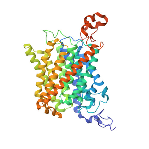
    Mep2 proteins are fungal transceptors that play an important role as ammonium sensors in fungal development. Mep2 activity is tightly regulated by phosphorylation, but how this is achieved at the molecular level is not clear. Here we report X-ray crystal structures of the Mep2 orthologues from Saccharomyces cerevisiae and Candida albicans and show that under nitrogen-sufficient conditions the transporters are not phosphorylated and present in closed, inactive conformations. Relative to the open bacterial ammonium transporters, non-phosphorylated Mep2 exhibits shifts in cytoplasmic loops and the C-terminal region (CTR) to occlude the cytoplasmic exit of the channel and to interact with His2 of the twin-His motif. The phosphorylation site in the CTR is solvent accessible and located in a negatively charged pocket ∼30 Å away from the channel exit. The crystal structure of phosphorylation-mimicking Mep2 variants from C. albicans show large conformational changes in a conserved and functionally important region of the CTR. The results allow us to propose a model for regulation of eukaryotic ammonium transport by phosphorylation.


  • Organizational Affiliation

    Institute for Cell and Molecular Biosciences, The Medical School, Newcastle University, Newcastle upon Tyne NE2 4HH, UK.


Macromolecules
Find similar proteins by:  (by identity cutoff)  |  3D Structure
Entity ID: 1
MoleculeChains Sequence LengthOrganismDetailsImage
MEP2486Candida albicansMutation(s): 0 
Membrane Entity: Yes 
UniProt
Find proteins for Q59UP8 (Candida albicans (strain SC5314 / ATCC MYA-2876))
Explore Q59UP8 
Go to UniProtKB:  Q59UP8
Entity Groups  
Sequence Clusters30% Identity50% Identity70% Identity90% Identity95% Identity100% Identity
UniProt GroupQ59UP8
Sequence Annotations
Expand
  • Reference Sequence
Experimental Data & Validation

Experimental Data

  • Method: X-RAY DIFFRACTION
  • Resolution: 1.47 Å
  • R-Value Free: 0.150 
  • R-Value Work: 0.137 
  • R-Value Observed: 0.138 
  • Space Group: H 3
Unit Cell:
Length ( Å )Angle ( ˚ )
a = 104.88α = 90
b = 104.88β = 90
c = 160.65γ = 120
Software Package:
Software NamePurpose
PHENIXrefinement
XDSdata reduction
Aimlessdata scaling
PHASERphasing

Structure Validation

View Full Validation Report


Ligand Structure Quality Assessment 


Entry History 

Deposition Data

Revision History  (Full details and data files)

  • Version 1.0: 2016-01-20
    Type: Initial release
  • Version 1.1: 2016-05-04
    Changes: Database references
  • Version 1.2: 2016-05-11
    Changes: Database references
  • Version 1.3: 2020-07-29
    Type: Remediation
    Reason: Carbohydrate remediation
    Changes: Advisory, Data collection, Derived calculations, Other, Structure summary
  • Version 1.4: 2024-01-10
    Changes: Advisory, Data collection, Database references, Refinement description, Structure summary