5AES

Crystal Structure of murine Chronophin (Pyridoxal Phosphate Phosphatase) in Complex with a PNP-derived Inhibitor


Experimental Data Snapshot

  • Method: X-RAY DIFFRACTION
  • Resolution: 2.75 Å
  • R-Value Free: 0.247 
  • R-Value Work: 0.190 
  • R-Value Observed: 0.192 

wwPDB Validation   3D Report Full Report


Ligand Structure Quality Assessment 


This is version 2.1 of the entry. See complete history


Literature

Synthesis of Hydrolysis-Resistant Pyridoxal 5'-Phosphate Analogs and Their Biochemical and X-Ray Crystallographic Characterization with the Pyridoxal Phosphatase Chronophin.

Knobloch, G.Jabari, N.Stadlbauer, S.Schindelin, H.Kohn, M.Gohla, A.

(2015) Bioorg Med Chem 23: 2819

  • DOI: https://doi.org/10.1016/j.bmc.2015.02.049
  • Primary Citation of Related Structures:  
    5AES

  • PubMed Abstract: 

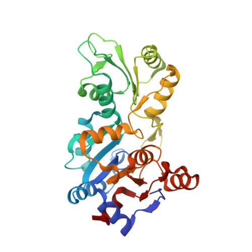
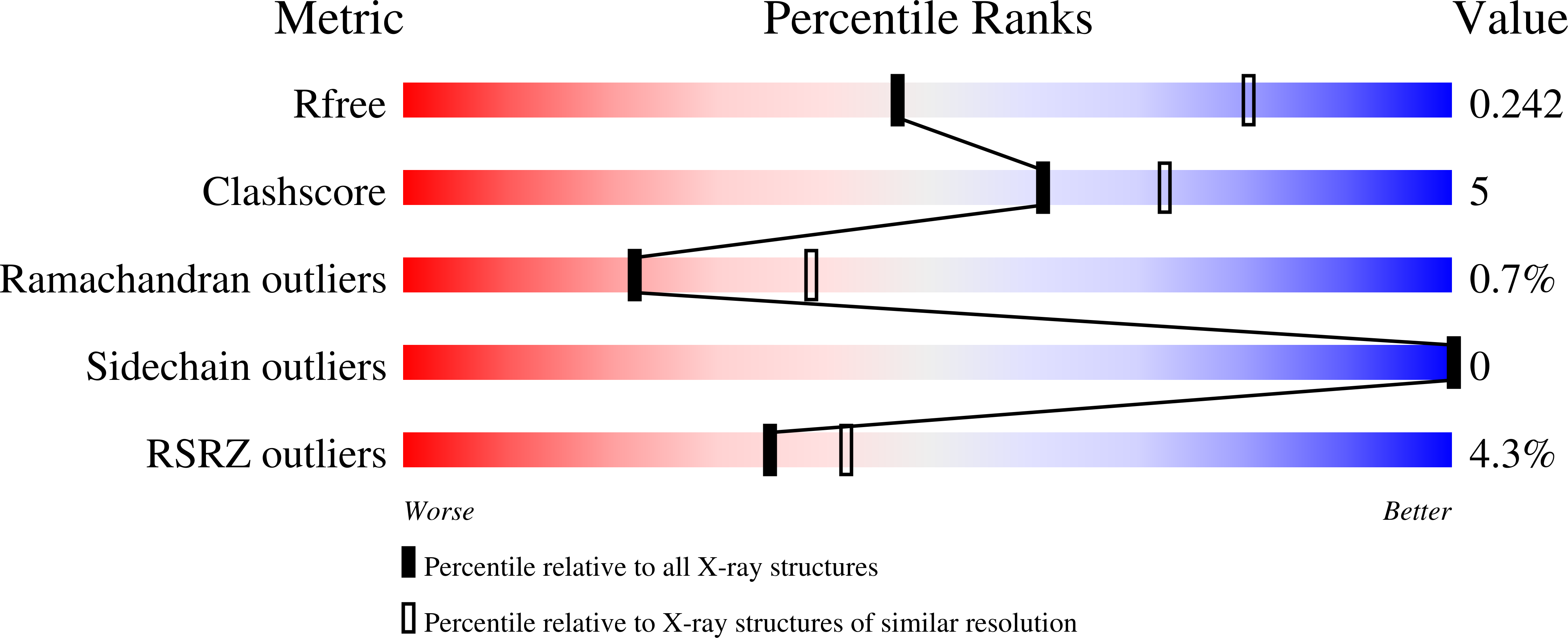
    A set of phosphonic acid derivatives (1-4) of pyridoxal 5'-phosphate (PLP) was synthesized and characterized biochemically using purified murine pyridoxal phosphatase (PDXP), also known as chronophin. The most promising compound 1 displayed primarily competitive PDXP inhibitory activity with an IC50 value of 79μM, which was in the range of the Km of the physiological substrate PLP. We also report the X-ray crystal structure of PDXP bound to compound 3, which we solved to 2.75Å resolution (PDB code 5AES). The co-crystal structure proves that compound 3 binds in the same orientation as PLP, and confirms the mode of inhibition to be competitive. Thus, we identify compound 1 as a PDXP phosphatase inhibitor. Our results suggest a strategy to design new, potent and selective PDXP inhibitors, which may be useful to increase the sensitivity of tumor cells to treatment with cytotoxic agents.


  • Organizational Affiliation

    Institute of Pharmacology and Toxicology, University of Würzburg, Versbacher Strasse 9, D-97078 Würzburg, Germany; Rudolf Virchow Center for Experimental Biomedicine, University of Würzburg, Josef-Schneider-Strasse 2, D-97080 Würzburg, Germany.


Macromolecules
Find similar proteins by:  (by identity cutoff)  |  3D Structure
Entity ID: 1
MoleculeChains Sequence LengthOrganismDetailsImage
PYRIDOXAL PHOSPHATE PHOSPHATASE
A, B
293Mus musculusMutation(s): 0 
EC: 3.1.3.74
UniProt & NIH Common Fund Data Resources
Find proteins for P60487 (Mus musculus)
Explore P60487 
Go to UniProtKB:  P60487
IMPC:  MGI:1919282
Entity Groups  
Sequence Clusters30% Identity50% Identity70% Identity90% Identity95% Identity100% Identity
UniProt GroupP60487
Sequence Annotations
Expand
  • Reference Sequence
Small Molecules
Experimental Data & Validation

Experimental Data

  • Method: X-RAY DIFFRACTION
  • Resolution: 2.75 Å
  • R-Value Free: 0.247 
  • R-Value Work: 0.190 
  • R-Value Observed: 0.192 
  • Space Group: I 2 3
Unit Cell:
Length ( Å )Angle ( ˚ )
a = 166.81α = 90
b = 166.81β = 90
c = 166.81γ = 90
Software Package:
Software NamePurpose
PHENIXrefinement
iMOSFLMdata reduction
SCALAdata scaling
PHASERphasing

Structure Validation

View Full Validation Report


Ligand Structure Quality Assessment 


Entry History 

Deposition Data

Revision History  (Full details and data files)

  • Version 1.0: 2015-04-01
    Type: Initial release
  • Version 1.1: 2015-04-15
    Changes: Atomic model, Other
  • Version 1.2: 2015-06-10
    Changes: Database references
  • Version 2.0: 2017-06-28
    Changes: Atomic model, Data collection
  • Version 2.1: 2024-01-10
    Changes: Data collection, Database references, Derived calculations, Other, Refinement description